-
E-mail
info@phenom-china.com
-
Phone
18516656178
-
Address
Room T5705, Shanghai Hongqiao Libao Plaza, No. 88 Shenbin Road, Hongqiao Town, Minhang District, Shanghai
Product Categories
Funa Scientific Instruments (Shanghai) Co., Ltd


Pharos STEM desktop biological scanning electron microscope
NegotiableUpdate on 12/28
- Model
- Nature of the Manufacturer
- Producers
- Product Category
- Place of Origin
Overview
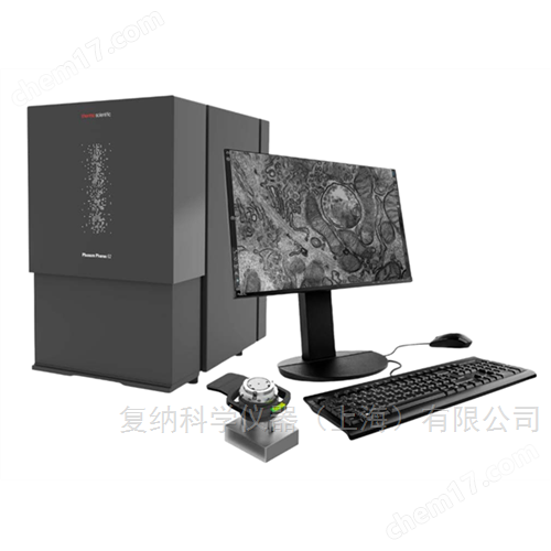
The Pharos STEM desktop biological scanning electron microscope is equipped with a STEM detector and utilizes a high brightness FEG electron source to image thin samples in transmission mode. The dedicated sample holder can easily load a conventional 3mm diameter transmission electron microscope (TEM) mesh, achieving fast and safe switching of samples. STEM detectors can provide bright field (BF), dark field (DF), and high angle annular dark field (HAADF) images, and support custom selection of imaging modes.
Product Details
Pharos STEM desktop biological scanning electron microscopeEquipped with STEM detectors and utilizing FEG high brightness electron sources, thin samples can be imaged in transmission mode. The dedicated sample holder can easily load a conventional 3mm diameter transmission electron microscope (TEM) mesh, achieving fast and safe switching of samples. STEM detectors can provide bright field (BF), dark field (DF), and high angle annular dark field (HAADF) images, and support custom selection of imaging modes.
Product Features:
1. High resolution:Using field emission filaments with a resolution better than 1 nm, all organelles related to diagnosis can be identified
2. 20KV low acceleration voltage:Low acceleration voltage brings better contrast and lower sample damage, which can be used to observe unstained biological samples and simplify your sample preparation process
3. Simple and fast operation:No need for professional operators, suitable for student operation, 15 second ultra fast vacuum pumping, 30 second imaging
4. Multiple imaging functions:BF,DF,HAADF, customize
5. Flexible installation:Desktop type electron microscope, compact in size, efficient in shock and magnetic resistance, no need to worry even when placed on high floors
6. Extremely low cost of ownership:No need for regular maintenance of the optical path
Application:
1. Pathological section
All organelles related to diagnosis can be identified, with a field of view 200 times that of traditional transmission electron microscopy, improving screening efficiency.
Left: ultrastructure of blood vessels, endothelial cells, and red blood cells; Right: Within myocardial cells: ultrastructure of myocardial fibers, mitochondria, and adipose tissue
2. Extracellular vesicles
The characterization of stereoscopy under low voltage has significant advantages and can be used for the observation of small-sized samples such as exosomes, viruses (AAV, tobacco mosaic virus), protein particles, plasmids, etc.
Left: TEM; Right: Pharos STEM
3. Undyed samples
Unpigmented samples can also be clearly visible, simplifying your sample preparation process and reducing sample shrinkage and heavy metal precipitation artifacts caused by staining.
Left: staining; Right: unstained
Specification parameters:
Pharos STEM desktop biological scanning electron microscope
Sample compatibility: ø 3 mm TEM mesh (fixture fixed)
Imaging time:<40 seconds*
Imaging modes: BF, DF, HAADF, custom**
Imaging workflow: fixed WD, settingsreasonableDetector with integrated UI
Vacuum degree: 0.1, 10&60 Pa
Resolution: ≤ 1 nm
*Time from loading the sample to presenting the image
**STEM has 11 segmented detectors, which users can customize and choose from
Similar Product Recommend