-
E-mail
info@phenom-china.com
-
Phone
18516656178
-
Address
Room T5705, Shanghai Hongqiao Libao Plaza, No. 88 Shenbin Road, Hongqiao Town, Minhang District, Shanghai
Product Categories
Funa Scientific Instruments (Shanghai) Co., Ltd
Feina desktop field emission scanning electron microscope | Research grade desktop electron microscope
NegotiableUpdate on 12/28
- Model
- Nature of the Manufacturer
- Producers
- Product Category
- Place of Origin
Overview
Feina desktop field emission scanning electron microscope | Scientific grade desktop electron microscope adopts a high brightness Schottky field emission electron source, with high-resolution, low-voltage imaging and integrated energy spectrum analysis functions, suitable for materials science, lithium batteries, semiconductors, biology and other fields. Easy to operate, no need for gold spraying, supports fast vacuum and automatic detection, combines scientific research grade performance with desktop level convenience, suitable for high-frequency use and various laboratory environments.
Product Details
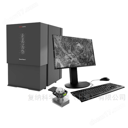
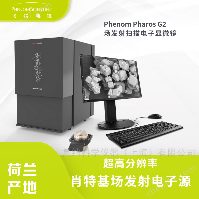
Phenom Pharos G2Feina desktop field emission scanning electron microscope | Research grade desktop electron microscope
Phenom Pharos G2It is the second generation launched by Funa in the NetherlandsSchottky field emission electron source desktop scanning electron microscope(Desktop field emission SEM). Equipment setBack scattered electron imaging (BSE)+secondary electron imaging (SE)+integrated energy dispersive spectroscopy (EDS) analysisIntegrating high resolution, high efficiency, and full automation for scientific research level microscopic characterization.
Thanks to the high brightness Schottky field emission electron source, Phenom Pharos G2Clear images can still be obtained under low voltage conditionsSignificantly reducing sample damage and electron beam penetration, very suitableInsulation materials, electron beam sensitive samples, biomaterials, polymersMicroscopic analysis in various fields.
Product Highlights | Optimal Selection of Research Grade Desktop Scanning Electron Microscopes
1. A true field emission level desktop electron microscope
adoptHeat field emission electron source (Schottky FEG)
High brightness, high stability, long lifespan, suitable for long-term scientific research experiments
2. High resolution imaging performance
Resolution better than1.5 nm
Fine surface structure can still be preserved in imaging under low voltage
More realistic restoration of sample morphology of nanomaterials, metals, semiconductors, polymers, etc
3. Multi mode imaging+energy spectrum integration
Backscattered Electron Imaging (BSE)
Secondary Electronic Imaging (SE)
Integrated energy dispersive spectroscopy (EDS) (high-speed stability)
One device can complete itMorphological and compositional analysis
4. Easy to operate and quick to get started
Automated design, 30 minutes to get started
Color optical microscope panoramic navigation for more intuitive positioning
integrationAutomatic Motor Sample StandNo threshold for operation
User Experience Upgrade | Suitable for Laboratories, R&D Centers, Production Quality Inspection
5. Fast, easy, and no need to spray gold
Built in vacuum lock,15 secondsVacuum extraction can be completed
Directly observe non-conductive samples without the need for metal coating treatment
More friendly to electron beam sensitive samples
6. High stability and earthquake resistant design
built-in27 sets of independent shock absorption modules
No need for additional seismic platforms, suitable for school laboratories, corporate R&D rooms, and small office areas
7. Maintenance free electronic optical path
No adjustment, no centering, no maintenance required
Ensure imaging consistency and significantly reduce maintenance difficulty
Low maintenance cost | Suitable for high-frequency use in scientific research and engineering scenarios
The Feina desktop scanning electron microscope is used for itsStable, durable, and highly automatedIts characteristics are widely used in research institutes, material research and development, lithium battery industry, semiconductor testing, quality analysis and other fields.
Long lifespan and stable performance of the filament
The electron microscope system is equipped with hardware security protection to avoid damage caused by misoperation
Free remote network diagnosis
-
Post maintenance is simple, and the total cost is significantly lower than that of traditional ground-based field emission electron microscopes

Applicable application areas
Materials Science (Metals, Ceramics, Polymers, Composite Materials)
Analysis of lithium battery materials and positive and negative electrode powders
Semiconductor, Microelectronics, and Wafer Defect Detection
Biomaterials, tissue structure, and micro/nano structure analysis
Surface engineering, coating, and film research
Industrial Quality Inspection and Failure Analysis
Why choose Phenom Pharos G2?
✔ Research grade imaging quality
✔ Desktop level volume, no need for computer room
✔ High degree of automation, zero threshold for operation
✔ Low maintenance cost and high reliability
✔ One machine completes morphology and element analysis
To obtainFeina desktop field emission scanning electron microscope | Research grade desktop electron microscopeTechnical parameters, configuration list, application cases or quotationsWelcome to contact us:
Similar Product Recommend