-
E-mail
info@phenom-china.com
-
Phone
18516656178
-
Address
Room T5705, Shanghai Hongqiao Libao Plaza, No. 88 Shenbin Road, Hongqiao Town, Minhang District, Shanghai
Product Categories
Funa Scientific Instruments (Shanghai) Co., Ltd
Feina Desktop Scanning Electron Microscope - Automated Quality Control Solution
NegotiableUpdate on 12/28
- Model
- Nature of the Manufacturer
- Producers
- Product Category
- Place of Origin
Overview
Phenom XL G3 Feina Desktop Scanning Electron Microscope - Automated Quality Control Solution is Thermo Scientific's flagship desktop scanning electron microscope platform, built on nearly 80 years of electron microscope technology accumulation from Philips FEI Thermo Fisher. It is stable, reliable, easy to deploy, and highly automated, designed specifically for production quality control, high-throughput testing, and research and development scenarios.
Product Details
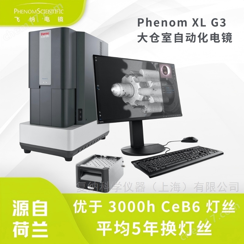
Feina Desktop Scanning Electron Microscope - Automated Quality Control Solution
Automated desktop scanning electron microscope designed for quality control and research and development
Phenom XL G3Feina Desktop Scanning Electron Microscope - Automated Quality Control SolutionIt is the flagship desktop scanning electron microscope platform under Thermo Scientific, built on nearly 80 years of electron microscope technology accumulation from Philips FEI Thermo Fisher Scientific. It is stable, reliable, easy to deploy, and highly automated, specifically designed forProduction quality control, high-throughput testing, and research and development scenariosAnd design.
Large warehouse design, capable of accommodating 36 samples
Complete sample installation to drawing in 1 minute
Automation, AI analysis, and energy spectrum integration
3000h CeB6 filament, replaced every 5 years on average
Suitable for various industries such as metals, electronic ceramics, semiconductors, powders, filter membranes, battery materials, etc.

⭐ Core Advantage (Strong Conversion Module)
01 | 80 years of electron microscopy technology inheritance, with over 5000 installed units in the entire (quan) sphere
From Philips Electron Microscopy Department to FEI, and then to Thermo Scientific, the Phenom series represents the leading standards in electron microscopy design and manufacturing.
In 1939Le Poole proposed the concept of electron microscopy
In 1944150kV electron microscope is introduced
In 1972The first Philips SEM
In 2006The first desktop SEM - Phenom was born
so far: Form a multi line product matrix of desktop CeB6/desktop field emission/industry-specific electron microscopy

02 | No need to renovate laboratory: use as you go, worry free and save money
Built in shock absorption system
Low environmental requirements, no need for darkroom or water cooling
Suitable for production workshops, QC laboratories, and high-rise office areas
Even supports in car and field use
I have participated in geological exploration missions such as Iceland's volcanic crater and Spain's uninhabited island, verifying their environmental reliability.


03 | CeB6 filament: 3000 hour lifespan, replaced every 5 years
High brightness cerium hexaboride (CeB6) filament
Replace once every 5 years on average, operate continuously 24/7 without pressure
Extremely low maintenance cost, suitable for production lines and long-term experimental use
Compared to ordinary metal filaments, which have a lifespan of 50-200 hours, the cost-effectiveness advantage is obvious.

04 | Low voltage imaging: Clear without gold spraying
Phenom XL G3 adopts a new vacuum structure and upgraded backscatter detector (BSD):
Higher signal-to-noise ratio at low voltage (2-5 kV)
No need to spray gold on non-conductive and sensitive samples
Improved imaging efficiency in automated processesAbout 30%
Applicable samples:Fiber, paper, coating, polymer, electronic ceramic CMOS、 Solar cells, etc.

Pyramid structure of solar cells
Magnification factor: 10000X
2kV, Unpainted gold, secondary electron image
You can clearly see the pyramid structure and details

electronic ceramics
Magnification factor: 1000X
5kV, Backscattered electron image
It is possible to observe the grain size and voids within the ceramic

CMOS detector
Magnification factor: 5000X
2kV, Secondary electron image
Clear visibility of pixel units and their surface pollutants

Fiber without gold spray under scanning electron microscope (top image), paper without gold spray (bottom image)
05 | Industry 4.0 Automation Platform: Supports AI&Python scripting
1 minute from sample loading to image output
Improved detection flux compared to traditional electron microscopy5-10 times.
Automated analysis software
ParticleMetric: Particle Statistics
FiberMetric: Fiber Size Analysis
-
Batch output standard reports to achieve true quantitative quality control

From "seeing" to "calculating": professional automated analysis software, specialized software such as ParticleMetric and Fiber Metric. Automated completion of statistical tasks such as particle size, morphology, and size distribution that require several hours of manual labor, outputting standardized reports, and achieving true quantitative quality control.
Open Programming Interface (PPI)
Support Python scripting
Can be integrated with production lines and LIMS systems
Support unmanned batch detection at night
-

Deep integration, on-demand ": Open automation interface, providing Python script control (PPI), can be seamlessly integrated into customers' existing automation production lines and LIMS systems. A precise automated motor table, combined with software scripts, can accommodate 36 samples at a time, creating an "unmanned quality inspection factory". Customized programs can automatically take photos and analyze large quantities of samples at night or during rest time, improving equipment utilization.
Large sample panoramic puzzle (MAPS)
View the whole picture with one image, analyze multiple points without omission
Automatically obtain panoramic high-definition images on large samples such as wafers, filter membranes, and metal fracture surfaces, and perform high magnification analysis on preset key points to ensure comprehensive detection.

06 | Integrated Spectral Design: A Software for Imaging and Elemental Analysis
No need to switch software, no data loss, easy training, and unified after-sales service.
ChemiSEM real-time color imaging
Synchronous acquisition of imaging and energy spectrum
Real time color element distribution display
-
Suitable for materials such as metals, ceramics, batteries, coatings, semiconductors, etc

ChemiSEM Color Imaging Technology
ChemiPhase Phase Phase Analysis
Automatic identification of physical phases
Output phase composition and area fraction
-
Especially friendly to complex materials such as battery cathodes, ceramics, metallurgy


ChemiPhase phase phase analysis software
Phase Mapping Phase Distribution Diagram
Based on pixel level quantitative element analysis
-
Automatically identify multiple material phases

Phenom MAPS large-area image stitching

Specifications
Imaging mode
Optical microscope: 3-16 ×
Electron microscope: 200000 ×
lighting system
CeB6 tube filament
Acceleration voltage: 2/5/10/15/20 kV (continuously adjustable)
Vacuum: low medium high mode
performance
Resolution: better than 8 nm
Vacuum extraction time: 30 seconds
Carrier platform
Large warehouse room, capable of supporting large-sized samples
Can accommodate up to 36 samples for automatic testing
Applicable Industries
Metal material analysis/metallurgical quality inspection
Battery and Material Research and Development
Electronic ceramics/semiconductors/components
Powder material screening
Filter membrane particle and fiber statistics
Surface research, coating analysis
Universities and research institutions
Why choose Phenom XL G3?
International brand technology inheritance (Philips → FEI → Thermo Fisher)
A true Industry 4.0 level automated desktop electron microscope
Support AI, Python, and unmanned detection
Complete spectrum and imaging integrated solution
Large sample, high flux quality control
-
On average, replacing the filament every 5 years results in extremely low operating costs
Similar Product Recommend