-
E-mail
info@phenom-china.com
-
Phone
18516656178
-
Address
Room T5705, Shanghai Hongqiao Libao Plaza, No. 88 Shenbin Road, Hongqiao Town, Minhang District, Shanghai
Product Categories
Funa Scientific Instruments (Shanghai) Co., Ltd
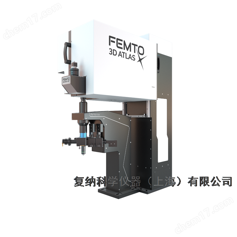
Femto3D Atlas acousto-optic three-dimensional two-photon microscope
NegotiableUpdate on 12/28
- Model
- Nature of the Manufacturer
- Producers
- Product Category
- Place of Origin
Overview
Femtronics is a company headquartered in Budapest, Hungary, specializing in neuroscience research and publishing over 200 top tier journal publications. Its star product, Femto3D Atlas acousto-optic three-dimensional two-photon microscope, adopts innovative acousto-optic scanning technology, providing ultra-high scanning speed and flexible imaging solutions, suitable for various scientific research fields. The product has a plug and play version and rich optional functions, supporting real-time motion correction and multimodal imaging, and assisting cutting-edge neuroscience research.
Product Details
Femtronics - At the forefront of neuroscience for 20 years
Femtronics is headquartered in Budapest, Hungary, and develops revolutionary technologies and cutting-edge tools for tens of thousands of neuroscience researchers worldwide. It has already createdMore than 20 world recordsThe Technical Achievements Publication Super200 copiesMost of the results have been published in top journals such as Nature, Science, Cell, and Neuron. Currently, its two-photon microscope is available worldwide45 of themNational installed capacity super150 unitsService exceeds300 usersCovering top international research institutions.
Femto3D Atlas acousto-optic three-dimensional two-photon microscope——Femtronics' flagship product
Femto3D Atlas acousto-optic three-dimensional two-photon microscopeAdopting innovative approachesSound and light scanning technologyIt can achieve ultra fast real-time in vivo 3D imaging with scanning speeds far higher than market products. 3D random access point scanning speed up to100 kHzThe high-speed arbitrary frame scanning speed is40 fps@510 ×510 pxWhen the target is focused on the field of view, up to3000 fps.
Integrated solution:With all existing laser scanning technologies, one device can meet your various imaging needs.
Ultra high speed 3D imaging:By adopting innovative sound and light technology, 3D imaging can be directly performed without moving the sample or objective lens, and the scanning speed of any point can reach100 kHz, capable of detecting ultrafast transients such as action potentials, and can reach up to40 fps@510 × 510 px, 500 × 500 µm2.
High flexibility:Equipped with flexible line, surface, and volume scanning capabilities, it can achieve arbitrary changes in shape, size, and direction in three-dimensional space3D network imaging with over 2000 neurons.
No scanner noise:Adopting sound and light scanning technology, without traditional scanner noise, it is more suitable for behavioral research.
Widely used:Deep functional imaging, network imaging, dendritic imaging, voltage imaging, calcium imaging, cage release, optogenetics, organoid imaging, electrophysiology, behavioral research, light stimulation, 2P-FLIM, blood flow research, developmental biology, plant research, etc.
Atlas acousto-optic 3D two-photon microscope (plug and play type)——Compact size, plug and play function
moreAtlas plug and play modelAvailable for selection, compact in size, plug and play,Within 1 hourComplete installation, can move freely, and flexibly adapt to laboratory needs.
Atlas acousto-optic 3D two-photon microscope - product selection
3D real-time motion correction:Provide real-time motion correction along the X, Y, and Z (axial) directions to effectively eliminate motion artifacts.
4D beam adjustment device:Used to control the beam and improve beam stability, it can not only eliminate many optical artifacts, but also prepare the beam for any specific application (such as deep functional imaging, etc.).
Dual laser source:Using two independent laser sources with different wavelengths in the same ATLAS scanning head can achieve quasi synchronous imaging and optical stimulation.
External expansion kit:Used for studying cells in a controlled environment in vitro, it can image acute brain slices or cultured cells and tissues.
Green lighting:The green illumination of LED light source can achieve high contrast visualization of blood vessels.
LED light source:It can uniformly stimulate molecules and cells throughout the entire field of view, and by combining this module with a gate detector, millisecond level switching between stimulation and imaging can be achieved.
Similar Product Recommend