-
E-mail
sende_services@outlook.com
-
Phone
17688459448
-
Address
No. 983 Huangpu Avenue East, Huangpu District, Guangzhou City, Guangdong Province
Product Categories
Guangdong Sende Instrument Co., Ltd

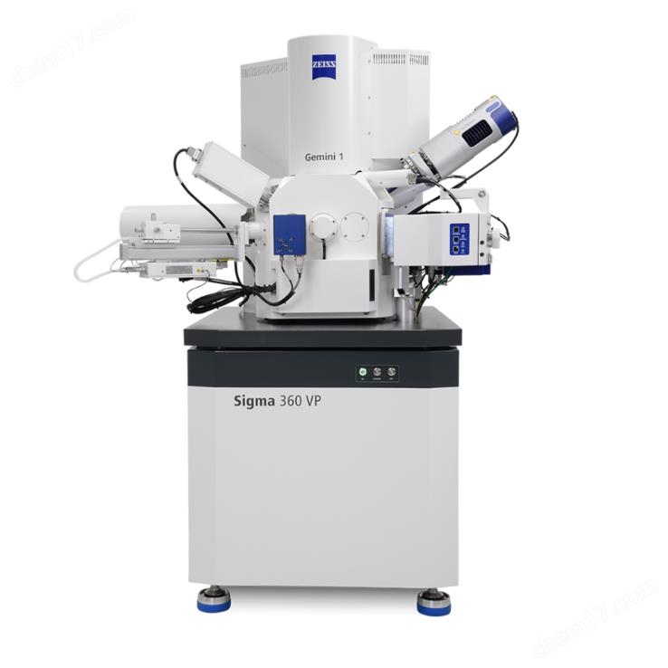
Ultra high speed scanning electron microscope
NegotiableUpdate on 02/08
- Model
- Nature of the Manufacturer
- Producers
- Product Category
- Place of Origin
Overview
The ultra high speed scanning electron microscope fully utilizes the collection speed of up to 91 parallel electron beams to image centimeter level samples with nanometer resolution. This unique scanning electron microscope is designed for continuous and reliable operation around the clock. By simply setting up a high-throughput data acquisition workflow, MultiSEM can automatically complete high contrast image acquisition.
Product Details
Zeiss MultiSEM
Ultra high speed scanning electron microscope
Fully utilize the collection speed of up to 91 parallel electron beams to image centimeter level samples with nanometer resolution. This unique scanning electron microscope is designed for continuous and reliable operation around the clock. By simply setting up a high-throughput data acquisition workflow, MultiSEM can automatically complete high contrast image acquisition.
lExtremely fast imaging speed
lAutomatic large-scale image acquisition
lNano level details under macro information
lLow noise, high contrast image


Multiple electron beams and detectors in parallel
MultiSEMUltra high speed scanning electron microscopeMultiple electron beams (green: illumination path) and detectors were used in parallel. Fine tuning the detection pathway (red) can collect a large amount of secondary electrons (SE) for imaging. The electron beams are arranged in a hexagonal pattern, and each electron beam performs a synchronous scanning program at a sample position to obtain a single sub image. Then, the entire image is generated by merging all images and stitching them together. Parallel computer setup programs are used to quickly record data to ensure high overall imaging speed. In the MultiSEM system, image acquisition and workflow control are independent.
Integrated workflow
Continuous slice tomography for collecting large volume samples

Automatic slicing
Use ATUMtome to automatically slice resin embedded biological tissues. Up to can be collected in one day1000A continuous slice.

Sample inlay
Insert the sliced tape onto the silicon wafer and image the sample using an optical microscope. Transfer the wafer to MultiSEM, use Overview to navigate and design your experiment.

Experimental setup
The entire experiment can be set up using a single graphical control center. By efficiently detecting and locking regions of interest through automatic slicing, time can be saved.
Application case of Zeiss MultiSEM


Similar Product Recommend