- Phone
-
Address
Room 3002, 3003, 3011, Dongshan Square, No. 69 Xianlie Middle Road, Yuexiu District, Guangzhou City
Product Categories
Fujifilm and Light (Guangzhou) Trading Co., Ltd

Local tissue structure fluorescent dye HistoBright
NegotiableUpdate on 05/06
- Model
- Nature of the Manufacturer
- Producers
- Product Category
- Place of Origin
Overview
HistoBright, a local tissue structure fluorescent dye, is an environmentally responsive membrane staining fluorescent pigment that can achieve two-photon excitation. It has excellent tissue permeability and can change fluorescence characteristics according to the local structure of the tissue, thus achieving high contrast tissue visualization. By combining tissue transparency treatment with two-photon laser microscopy, tissue blocks can be observed in depth and analyzed for spatial structure.
Product Details
Fluorescent dyes capable of visualizing local tissue structures
HistoBright
HistoBright is an environmentally responsive membrane staining fluorescent pigment that can achieve two-photon excitation. It has excellent tissue permeability and can change its fluorescence characteristics according to the local structure of the tissue, thus enabling high contrast tissue visualization. By combining tissue transparency treatment with two-photon laser microscopy, tissue blocks can be observed in depth and analyzed for spatial structure.
※This product is based on the research results of Kochi University and Ehime University, and is produced and sold by Funakoshi Co., Ltd.
※This product is for research purposes only and cannot be used for purposes other than research.

HistoBright is an environmentally responsive fluorescent dye that can display a wide range of wavelengths based on the polarity of the solvent. When applied to tissues that have undergone transparency treatment, fluorescence changes in response to the microenvironment within the tissue, and is then combined with dual lightThe sub laser microscope can clearly observe the tissue structure.
Traditional organizational structure analysis methods and new three-dimensional organizational structure analysis
Traditional organizational structure analysis is achieved by staining tissues with colored dyes, such as hematoxylin eosin (HE) staining method. However, it is difficult to dye the entire tissue block with these colored dyes, and it is generally necessary to make thin slices before observation. However, thin slices can easily lose the three-dimensional information of tissue structure, so a large number of slices need to be prepared to reproduce the three-dimensional structure.
HistoBright is a novel environmentally responsive fluorescent dye synthesized by Dr. Yokosuke Renzi from the Faculty of Education and Research at Kochi University (the compound in the original paper is named PC). Currently, it is being developed and applied as a fluorescent imaging dye for three-dimensional tissue structures by Dr. Kenji Imamura, Dr. Masaki Murakami, and Dr. Ryosuke Kawakami from the Faculty of Medicine at Ehime University. HistoBright aggregates on biofilms and emits fluorescence depending on the local environmentGreen Light~near-infrared lightLarge scale changes. Therefore, when HistoBright is applied to biological tissues, it can induce changes in fluorescence tone based on tissue structure, thereby achieving high contrast observation of tissue structure. Due to the ability of HistoBright to perform two-photon excitation, in addition to confocal laser microscopy, two-photon laser microscopy can also be used for observation. In addition, in conjunction with tissue transparency treatment (except for the method of degreasing to maintain the membrane structure), HistoBright can be used to observe thick tissue samples without damaging the tissue structure (without preparing thin slices), and depth imaging can be performed through two-photon laser microscopy to achieve three-dimensional structural analysis of the tissue.
◆ Characteristics
An environmentally responsive membrane staining reagent that can exhibit changes based on the surrounding environmentGreen Light~near-infrared lightFluorescence characteristics. By distinguishing the fluorescence wavelength range that needs to be detected, high contrast local tissue structures can be achieved
Visualize the structure.
●Combined with non degreased tissue transparency treatments (such as RapiClear (SunJin Lab), LUCID, etc.), deep three-dimensional analysis of tissue structure can be achieved.
●The use of surfactants such as Triton X-100 and Tween 20 can dissolve lipid membranes in tissues, which may affect the staining of HistoBright. Therefore, it is not recommended to use them. If necessary, please discuss the applicable conditions.
●Observation can be performed using a confocal laser microscope (recommended excitation light: 488 nm laser) or a two-photon laser microscope (recommended excitation light: 960 nm laser).
●When using a two-photon laser microscope for observation, second harmonic generation (SHG) imaging can be combined to observe collagen fibers in unstained tissues while using this reagent.
●After staining and observation with HistoBright, HE staining can be performed.
◆ Original text
Inoue, K.,et al., J. Mater. Chem.B.,10(10), 1641~1649 (2022). Synthesis and photophysical properties of a new push-pull pyrene dye with green-to-far-red emission and its application to human cellular and skin tissue imaging. [PMID:35194628]
◆ Principle
Excitation/emission spectra
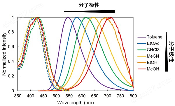
HistoBright is a solvent polarity responsive fluorescent dye that changes its fluorescence properties based on the molecular polarity of the solvent. According to the polarity of the solvent, fluorescence will shift fromgreen(Low polarity) towardsnear-infrared lightSignificant changes within the range of high polarity.
solvent |
Maximum absorption wavelength (nm) |
Maximum fluorescence wavelength (nm) |
Fluorescence quantum yield |
Toluene |
424 |
546 |
0.82 |
EtOAc |
418 |
581 |
0.77 |
CHCl3 |
427 |
610 |
0.83 |
MeCN |
414 |
641 |
0.65 |
EtOH |
427 |
691 |
0.2 |
MeOH |
426 |
712 |
0.07 |
Changes in fluorescence spectra in liposome membrane model
HistoBright exhibits high affinity for lipid membranes and displays different fluorescence characteristics based on the phase of the lipid membrane. Unlike the maximum fluorescence in the ordered phase Lo model (sphingomyelin (SM)/bile solid alcohol (Chol)) liposomes, the maximum wavelength in the disordered phase Ld model (DOPC) liposomes also shifted to a long wavelength range of about 30 nm, and the fluorescence quantum yield decreased from 0.72 (SM/Chol) to 0.37 (DOPC).
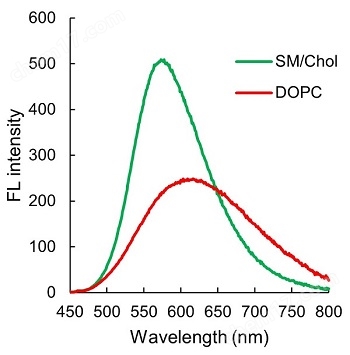
The above fluorescence spectrum data were provided by Dr. Yokosuke Renzi from Kochi University.
◆ Application data
Imaging of Transparent Normal Human Skin Tissue Using a Two Photon Laser Microscope
After fixing human normal skin tissue blocks with 4% paraformaldehyde/PBS, 500 μ m sections were prepared and subjected to tissue transparency treatment (LUCID) and HistoBright (10 μ M) staining for 76 hours. By using a two-photon laser microscope, tissue blocks were excited by laser at 960 nm, and then at 492 nm (SHG:cyan)、500-550 nm(green)、560~593nm(orange)And 593-690 nm(red)Obtain fluorescence images separately and create the following 4 composite images.
※ 492 nm(cyan)SHG signals derived from collagen fibers in the tissue, rather than fluorescence signals derived from HistoBright.
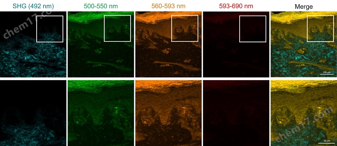
Next, use a two-photon laser microscope to continuously capture 500 μ m images at intervals of 5 μ m in the Z-axis direction, and use image analysis software for three-dimensional construction to visualize the fine structure of human skin tissue in a three-dimensional manner.
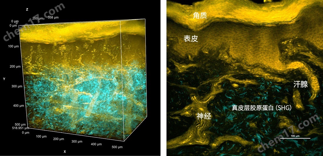
Three dimensional imaging of transparent human normal skin tissue using two-photon laser microscopy
After fixing human normal skin tissue blocks with 4% paraformaldehyde/PBS, 1mm slices were prepared and subjected to 72 hours of tissue transparency treatment (RapiClear, SunJin Lab) *, HistoBright (10 μ M) staining, and nuclear staining (Hoechst33342). By using a two-photon laser microscope, tissue blocks were excited by laser at 1100 nm, respectively, at<492 nm (SHG:cyan)、500~550 nm(green)、560~593 nm(orange)And 593-690 nm(red)Obtain fluorescence images separately and create the following 4 composite images. Based on the above conditions, take continuous shots at intervals of 5 μ m in the Z-axis direction, use image analysis software for 3D construction, and create animations from the continuous data.
The tissue blocks in this experiment were fixed without membrane permeation treatment, and were directly subjected to transparency treatment and staining.
Observation image of genuine leather |
Observation image of epidermis |
|
|

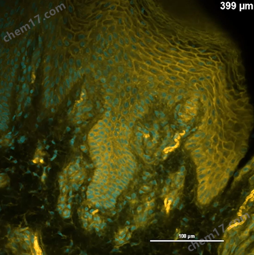
Please refer to labchem.fujifilm-wako.com.cn/product/show/1646. html for the complete video
Repeatedly used for HE staining
Stain with HistoBright and observe normal human skin cell tissue blocks, let them stand in PBS, and remove the transparency reagent. Then prepare thin slices and perform HE staining. After confirming HistoBright staining, clear HE staining can still be performed.
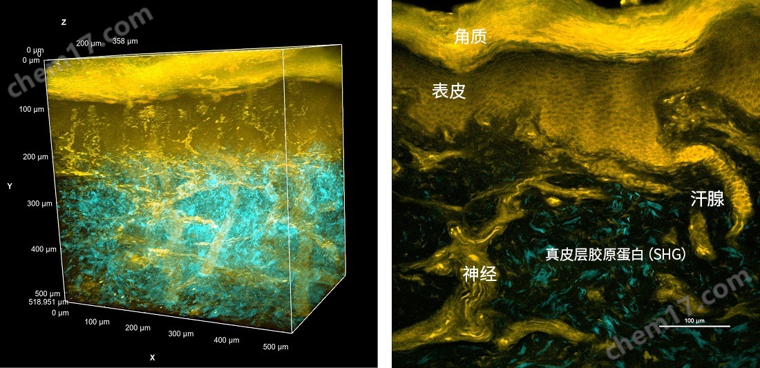
Observing staining under confocal laser microscopy and two-photon laser microscopy
After fixing human normal skin tissue blocks with 4% paraformaldehyde/PBS, 0.5 mm sections were prepared and subjected to 72 hours of tissue transparency treatment (RapiClear, SunJin Lab) and HistoBright (10 μ M) staining. Subsequently, observations were made using a confocal laser microscope (left image: excitation light 488 nm laser) and a two-photon laser microscope (right image: excitation light 960 nm laser). Although there is no significant difference between the two images, the SHG signal (collagen fibers) excited by two-photon excitation can be observed under a two-photon laser microscope.
The tissue blocks in this experiment were fixed without membrane permeation treatment, and were directly subjected to transparency treatment and staining.

The above image data were provided by Dr. Ryosuke Kawakami from Ehime University.
◆ Product List
| Product Number | Product Name | Product Specifications |
| FDV-0051 |
HistoBright |
0.1 mg |
Similar Product Recommend