- Phone
-
Address
Building 5, No. 3701 Xiang'an North Road, Xiamen Torch High tech Zone (Xiang'an) Industrial Zone
Product Categories
Xiamen Moji Biotechnology Co., Ltd

Mogengel® Cell culture plate (15 well)
NegotiableUpdate on 05/06
- Model
- Nature of the Manufacturer
- Producers
- Product Category
- Place of Origin
Overview
Mogengel Cell tube culture plate (15 well) is a high-quality consumable series product specifically designed for model based biological tube experiments, abbreviated as 15 well tube culture plate, suitable for forming tubular structures outside of cells.
Product Details
Mogengel®Cell tube culture plate(15Kong)
|
1 Product Description
Mogengel®Cell tube culture plate(15Kong)It is the abbreviation for the series of high-quality consumables specifically designed for the tube forming experiment of modular biology15Kongcheng tube culture plate is suitable for forming tubular structures in vitro.
2 Product Information
| Product Name | Product Item Number | specification | storage/transportation | shelf life |
| Mogengel®Cell tube culture plate(15Kong) | MS-TBFMW15 | 1branch/package | Room temperature storage | 60month |
3 intended use
Mogengel®Cell tube culture plate(15Kong)Used for endothelial cells or tumor cells in vitro3DenvironmentECM)A tubular cavity is formed in the middle to study the effects of certain genes or drugs on this process. Based on the theory that tumor growth and metastasis depend on angiogenesis, this experiment is also used to study the mechanism of tumor occurrence. This type of research is conducted in the process of tumor research, but the cost is expensive. This product aims to reduce the amount of reagents and cells used in tube experiments and lower the cost of scientific research.
Attention: Always wear when using, and follow safe laboratory procedures when handling substances such as human cells or other organisms and harmful substances.
Only for scientific research purposes.
4 Instructions for use: Organoid culture
a) Experimental supplies
Mogengel®Cell tube culture plate(15Kong)TheMogengel®MatrixStandard matrix adhesive (item number:0827045)Required cells and culture medium;
b) Experimental supplies
b.1, gel
Add to the lower chamber of the culture plate8-10MLMatrix adhesive, be careful not to produce bubbles. placed in37℃ gel in cell incubator5-15min.
b.2Cell experiments
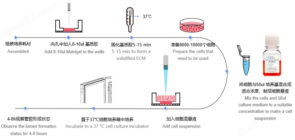
After pre starvation or direct digestion of different cells to obtain the expected state of cells, resuspend and dilute them in cell culture medium to the corresponding number of cells, and extract them50MLjoinMogengel®Cell tube culture plate(15Kong)In the middle, placed37Placement of ℃ incubator4-8hPre experiments can observe and confirm the experimental parameters of relevant cells at certain intervals.
watch1 HUVECCell usage for vascular formation experiment
| Number of cells (pcs) | 1*104 |
| The amount of cell culture medium used (μ g/mL)L) | 50ML |
b.3Cell experiments
Use an intelligent microscope system to capture the process of tracking tube formation, initiate real-time intelligent tube formation data analysis, and export the data. If there are no relevant devices, you can proceed to the steps according to the experimental requirements“b.4”.
b.4Cell experiments
1)Dye dilution: Use serum-free culture medium (such asDMEM)Diluted Calcein(12.5 µl calcein stock 1 µg/µl)The final concentration is6.25 µg/ml;
2)Remove the culture medium from the upper well and add10MlCalcein diluent;
3)Incubate at room temperature in the dark30After minutes, use sterilePBSWash three times;
4)Use485 nm/ 529 nmPerform immunofluorescence imaging on the channel.
b.5Image acquisition
After staining, use2The formation status of the magnifying glass collection tube (or directly collecting bright field images) is analyzed using image analysis software to analyze parameters such as tube area, tube length, tube quantity, ring area, ring quantity, and bud quantity for subsequent data analysis.
Similar Product Recommend