-
E-mail
sales@glo-bio.com.cn
-
Phone
13501678399
-
Address
Room 207A, Building L1-A, Hongqiao World Center, Lane 1588 Zhuguang Road, Qingpu District, Shanghai
Product Categories
Suzhou Grobel Biotechnology Co., Ltd


Miniscope Miniature Monochromatic Deep Brain Microscope System
NegotiableUpdate on 01/01
- Model
- Nature of the Manufacturer
- Producers
- Product Category
- Place of Origin
Overview
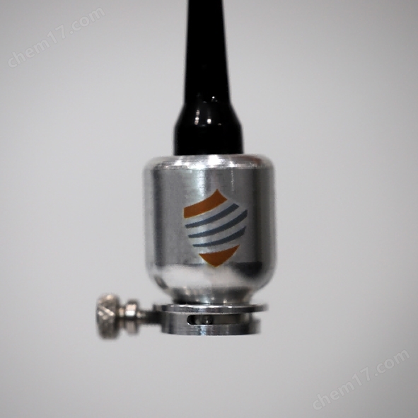
The Miniscope Micro Monochromatic Deep Brain Microscope System (Electronic Focusing Edition) uses a self focusing (GRIN) lens to image deep brain regions. The microscope body is connected to the imaging sleeve through threads (no tools required).
Product Details
Miniscope Micro Monochrome Deep Brain Microscope System (Electronic Focusing Edition)
Miniscope Miniature Monochromatic Deep Brain Microscope SystemUse a self focusing (GRIN) lens to image deep brain regions. The microscope body is connected to the imaging sleeve through threads (without the need for tools), and an optional optogenetics module can be used to regulate neuronal activity within the field of view or upstream and downstream. The optogenetics module uses an external light source without increasing the weight of the head. The 3rd generation eFocus fluorescence microscope can perform single-cell resolution imaging on freely moving animals, with an imaging field of view increased to 650 x 650 µ m and an electronic focus adjustment depth of 300 µ m. The more sensitive CMOS sensor allows the system to operate with less fluorescence excitation light, achieving high-quality imaging results and reducing photobleaching and phototoxicity, resulting in longer continuous acquisition time.
Electronic Focusing Imaging System - eTFMS3
Main features:
1) Electronic focusing: Software controlled electric focusing, no need for manual rotation adjustment, stable field of view, avoiding manual focusing stimulation of animals;
2) High sensitivity sensors require less fluorescence excitation light, reducing photobleaching and phototoxicity;
3) Simple connection: The microscope body is threaded to the imaging sleeve without the need for any tools;
4) Free movement: Equipped with Doric's high-quality commutator equipment, it can move in various behavioral operation boxes, mazes, and open fields;
5) At single-cell resolution, it can simultaneously capture the activity of over 1000 neurons;
6) Long term tracking of cells can last for several months;
7) Slow heating: External light source reduces the heat generated by the microscope;
8) Modular design, easy to carry and maintain.
System components:
Microscope driver (including LED light source), electron focusing microscope, imaging sleeve, commutator, gripper, simulation microscope, software, cable kit, etc.

Electronic Focusing Imaging System Combining Optogenetics - eTOSMS3
Electronic focusing+calcium ion imaging+optogenetics integrated system
System composition:
LISER ™ +LED (470nm) light source and driver, electron focusing microscope, imaging sleeve, commutator, microscope stand, virtual microscope, software, cable kit, etc
Main features:
1) Ca2+imaging and light stimulation (excitation/inhibition) can be performed simultaneously
2) Calcium ion probe+optogenetic opsin
3) Minimize biological interference and maximize signal-to-noise ratio
4) Synchronized tracking and behavioral analysis
Partial published articles
![]()
标 题: Drd3 Signaling in the Lateral Septum MediatesEarly Life Stress-Induced Social Dysfunction
Nucleus: Side Diaphragm (0.85, 0.35, 3.2)
Communication: Byung Kook Lim
Unit: UCSD
Direction: Social Behavior
Model: Snap in Model L, Doric Neuroscience Studio
Application: GCaMP6f
Using in vivo calcium imaging technology and a series of behavioral experiments, the team found that in mice that experienced early stress (3 hours of solitude per day within 14 days after birth),The Drd3 neurons in the lateral septum exhibit sluggish activity in response to social stimuli (when interacting with other mice).

![]()
Title: A Synaptic Threshold Mechanism for Computingescape decisions
Nucleus: Medial superior thalamus mSC (-0.2 to -0.5,+0.25, -2.2) dorsal sideGray matter dPAG around the aqueduct (-0.4 to -0.6,+0.25, -2.2)
Communication: Tiago Branco
Unit: UCL
Direction: Neural circuit of escape behavior
Model: Snap in Model L
Application: GCaMP6s


DPAG → Escape mSC Excitement → Activation
Similar Product Recommend