- Phone
-
Address
Room 405, Building B, No. 11 Xiyuan Eighth Road, Xihu Science and Technology Park, Hangzhou City
Product Categories
Hangzhou Xunshu Technology Co., Ltd

Micronucleus analysis/colony counting/cell counting combined instrument
NegotiableUpdate on 05/06
- Model
- Nature of the Manufacturer
- Producers
- Product Category
- Place of Origin
Overview
GenTox 3 consists of an Olympus CX31 microscope, microscope camera, micronucleus analysis software, MIC cell analysis software, and colony analysis system. It has functions such as intelligent analysis of red blood cell micronuclei, cell counting, and automatic colony counting. It is a high-performance imaging analysis tool for genetic toxicology.
Product Details
GenTox 3Micronucleus analysis/colony counting/cell counting combined instrument
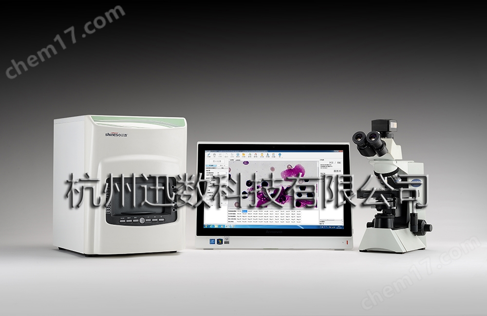
GenTox 3 consists of an Olympus CX31 microscope, microscope camera, micronucleus analysis software, MIC cell analysis software, and colony analysis system. It has functions such as intelligent analysis of red blood cell micronuclei, cell counting, and automatic colony counting. It is a high-performance imaging analysis tool for genetic toxicology.
Intelligent analysis system for red blood cell micronucleus
Realistic and delicate display of color cell images
Using an Olympus CX31 microscope and a 2/3-inch high-sensitivity CCD image sensor, quickly obtain bone marrow smears stained with Giemsa, with delicate images and high cell clarity.


Automatic calculation of micronucleus cell rate
Through deep learning of PCE and NCE cells and random resonance image processing, the proportion of PCE in total red blood cells was determined within 20 seconds; Within 60 seconds, 2000 PCE cells were captured from 200 different field of view micrographs, and the micronucleus cell rate was automatically identified and calculated, greatly improving the efficiency of microscopic examination.
Image database management, convenient human-machine operation
1. Review and retest: For identified cells, quick follow-up and localization can be carried out to retrieve the original images and coordinates.
2. Quickly establish a sample image library: five groups (high, medium, and low dose groups, positive and negative control groups), 50 glass slides, and 10000 photos (one micrograph represents one field of view) for easy retrieval at any time.


Dual-mode microscopic measurement
The digital micrometer dragged by the mouse can accurately measure the diameter, angle, curvature, circumference, area, etc. of cells. When the cell contour is clear and discretely distributed, automatic measurement mode can be used to automatically measure the measurement area, circumference, diameter, and roundness of all cells in just 1 second.
cell counting
Multi functional counting module, which can be used for cloning counting on porous plates and automatic counting of microscopic cells.
Blurred image clarity
27 image processing functions including adaptive enhancement, edge sharpening, background smoothing, filtering, edge detection, morphological operations, etc., enable clearer display of chromosome nuclear shape and more subtle observation of changes in chromosome number and structure.
Colony counting system
Fully enclosed scene lighting, high pixel camera, highlighting tiny bacterial colonies
Three color LED mixed soft light box, Crystal Sharp suspended dark field illumination, high fidelity fixed focus lens, 16 megapixel camera, sharp display of colony color and contour, providing image assurance for Ames test accuracy.
Security architecture design to achieve effective audit tracking
The four tiered system architecture of "system administrator, data administrator, operator, and reviewer" is divided into functions and permissions to ensure the security, integrity, and authenticity of data information.
System administrator: responsible for creating and managing accounts for all operators and auditors And login password.
And login password.Data administrator: responsible for archive management, sealing, and computer database management of all test data.
Operator: Responsible for testing, self checking, correcting, generating electronic reports, submitting for review, and printing reports on approved documents for culturing dish colonies
Auditor: Responsible for reviewing the test reports submitted by operators.
One click intelligent six modes
To facilitate rapid and accurate colony counting by experimenters, six intelligent modes have been optimized and designed based on common plate types:Flat sense mode, three-dimensional sense mode, small colony priority, large colony priority, same color bacteria priority, medium exclusion modeComplete colony counting with just one click.
Main functions and technical indicators of the instrument
1、 System composition
1 GenTox 3 host
Intelligent analysis software for red blood cell micronuclei; MIC analysis software; Colony analysis software; 1 encryptor
Lenovo all-in-one computer (nationwide warranty): dual core CPU/4G memory/1T hard drive/21.5 'color display, Windows 7 or Windows 10
Professional microscope camera, C-type adapter
Olympus CX-31 microscope
2、 Microscope parameters
Optical system: UIS2 optical system (infinity correction system);
Observation tube: The tilt angle of the tube is 30 degrees, the pupil spacing is 48-75mm, and the optical path selection is (50 binocular/50 camera)
Focusing: The vertical movement of the stage is guided by a roller (rack and pinion) mechanism, using coarse and fine coaxial knobs. The coarse adjustment stroke is 36.8mm per turn, the total stroke is 25mm, and the fine adjustment stroke is 0.2mm per turn. It is equipped with a coarse adjustment limiter and a tension adjustment ring;
Focusing lens: Abbe focusing lens, built-in daylight filter, numerical aperture 1.25 (when immersed in oil), built-in aperture stop;
Lighting system: Built in transmitted light Kohler lighting, 6V30W halogen lamp; 100-120V/220-240Vg 0.85/0.45A 50/60Hz;
Objective lens turntable: a fixed 4-hole objective lens turntable with a converter tilted inward;
Stage size: 188 × 134mm, range of motion: X-axis 76 × Y-axis 50mm, dual specimen holder;
Eyepiece: Field of view F.N. 20
Objective lens: flat field achromatic
4× N.A.0.1, W.D. 18.5mm
10× N.A.0.25 W.D. 10.5mm
40× N.A.0.65 W.D. 0.56mm
100× N.A.1.25 W.D. 0.13mm
3、 CCD camera parameters
Research grade color CCD large array camera
Sensor model/size: Sony ExView HAD CCD chip 1.4M/ICX285AQ (C); 2/3 inch
Pixel: 6.45x6.45 μ m
G light sensitivity, dark current: 1240mV with 1/30s; 10mv with 1/30s
FPS/Resolution: 15@1360x1024
Exposure time: 0.12ms~240s
Data interface: USB 2.0
4、 Micronucleus analysis software
1. Fast image acquisition
CCD connection: enables real-time dynamic observation of ultra large field of view microscopic images, reducing the amount of images captured.
CCD adjustment: It has the functions of adjusting exposure time and white balance
CCD photography: Microscopic image acquisition, automatic saving of batch images
2. Cell feature learning
Positive red blood cell learning: Randomly selecting typical mature red blood cells (NCE), intelligent learning, and memory cell characteristics
Polychromatic red blood cell learning: Randomly selecting typical immature red blood cells (PCE), intelligent learning, and memory cell characteristics
Revise selected cells: with undo, clear and reselect functions
3. Experimental parameter settings:Total number of observed red blood cells, number of observed polychromatic red blood cells
4. Analyze parameter adjustment:Resonance total intensity, affinity diffusion, micronucleus sensitivity
5. PCE and NCE analysis:Complete automatic recognition and capture of PCE and NCE in 20 seconds; Automatically calculate PCE/RBC
6. Micronucleus analysis:Complete PCE capture and intelligent recognition of micronuclei containing cells in 60 seconds; Automatic calculation of micronucleus cell rate
7. Information tracing:The detected PCE cell array is digitally located, and images and coordinates are recorded for follow-up verification of cell recognition accuracy
8. Data management:
Electronic records: record the operator's experimental data to ensure data accessibility and integrity;
Report output: Output in "PDF" or "EXCEL" format, the output report data is consistent with electronic records and cannot be changed.
Account management: graded management by administrators and operators, only authorized personnel can log in; Administrators comprehensively manage operator accounts, passwords, account freezing, etc.
Audit tracking: Record personnel identities, operational processes of each operator, including time, samples, whether statistical results have been modified, whether historical data has been deleted, and all historical records.
5、 MIC Microscopic Analysis Software
1. Image display and conversion
Image display: Real time dynamic observation, capturing any field of view image at any time
Image observation: with functions of rotation, enlargement, reduction, mirror conversion, and local observation
Image editing: It has functions such as cutting, copying, pasting, and text input for any area of the image
2. Microscopic image processing
Adaptive enhancement: By performing resolution enhancement processing on the original image that matches its features, the image becomes clearer and the edges are more prominent, facilitating the observation and recognition of subtle structures in the image.
Image adjustment: arbitrary adjustment of image brightness, contrast, saturation, RGB three colors, conversion of grayscale image and negative phase diagram
Image compensation: Various mathematical methods such as linear compensation, logarithmic compensation, Bell compensation, etc. are used to compensate for the distorted parts of the image, making the image clearer.
Image sharpening: By enhancing the high-frequency components of an image, the edges of the image become clearer.
Image smoothing: Through image smoothing processing, the background of the image is made uniform.
Image filtering: Six filtering methods, including Gaussian filtering, low-pass filtering, and median filtering, effectively improve image clarity.
Edge detection: Two detection methods and three operators combined with multiple detection options can more accurately extract image contours.
Morphological processing: Nonlinear mathematical morphological processing such as corrosion, expansion, opening, and closing.
3. Target measurement
Calibration: It has the function of online calibration of the system to achieve accurate measurement (default calibration values are built into the system)
Measurement function: online measurement of particle diameter, length, curvature, angle, any curve, area, etc
4. Particle statistics
Automatic statistics: Automatically count particles and display morphological parameters such as area, perimeter, diameter, roundness, etc. of each particle
Regional statistics: Any shape of region such as rectangle, circle, umbrella, etc. can be selected for statistics
Diameter classification statistics: Set the diameter range and count particles of specific sizes
Color recognition statistics: Select specific particles based on chromaticity, brightness, and saturation
Mouse click statistics: Adding or deleting particles with mouse clicks is convenient and fast
Adhesive segmentation processing: According to user needs, particles that adhere to each other can be automatically or manually segmented
Multiple statistical algorithms: using multiple segmentation algorithms, suitable for particle statistics with different backgrounds
Diversified book statistics: comprehensive statistics of multiple microscopic images
Automatic parameter conversion: Based on the statistical area and sample dilution, achieve automatic conversion
5. Drawing and Annotation
Drawing: You can draw lines, rectangles, circles, and any curves as needed for the opened image
Text editing: Text editing of opened images
Annotation: Convenient for annotating lines and angles
6. Report printing
Online editing: provides report writing templates, text input, and print preview
Report printing: automatic printing of images and statistical data
6、 Colony digital imaging host
1. Light source
Visible light: High brightness three color LED structured light
254nm UV: used for cavity disinfection
2. Light path and lighting control
Fully enclosed dark box: eliminating environmental stray light interference
Upper light source: Scene style 360 ° flexible shadowless lighting
Lower light source: Crystal Sharp floating dark field illumination
Up light, down light, dual light, UV, freely switchable, adjustable light intensity
3. Optoelectronic conversion
High definition industrial fixed focus lens: focal length 8mm, resolution 150lp/mm
Professional CMOS camera: 1/2.33-inch color CMOS sensor, 16 million pixels C-Mount、USB3.0
7、 Colony counting software
1. Basic colony counting function
Type of petri dish: pouring, coating, membrane filtration, spiral petri dish, 3M paper sheet, porous plate
One click intelligent counting (6 modes): flat sensing mode, three-dimensional sensing mode, small colony priority, large colony priority, same color bacteria priority, culture medium removal mode
Whole dish colony count: Count the total number of colonies and display them classified by 25 sizes
Region selection statistics: You can choose circular, rectangular, or arbitrarily delineated regions for statistics
Diameter classification statistics: Set a diameter range and count colonies of specific sizes
Mouse click statistics: Quickly mark and add colonies, suitable for counting colonies at the edge of culture dishes
Colony adhesion segmentation: Automatically segment colonies that adhere to each other, and users can choose to segment or not segment chain colonies
2. Advanced colony counting function
Dynamic adjustment statistics: The statistical results can be dynamically adjusted and corrected to quickly obtain the best statistical effect.
Deviation estimation statistics: suitable for situations with multiple and complex colony colors.
Level set multi model algorithm: search operation, obtain the best image segmentation effect, adapt to background changes in the culture medium
Specific colony statistics: Identify specific colonies based on their color, size, and contour characteristics
Trans statistics: suitable for extremely complex colony types and uniform culture medium background
3. Advanced tools
Manual counting correction: adding or deleting colonies
Exclude contaminated areas: Use the mouse to outline any contaminated area and automatically remove the bacterial count from the contaminated area
Background text elimination: automatically eliminates marker interference
Artificial Adhesive Segmentation: Manually segmenting multiple adhesive colonies
Automatic parameter conversion: Input the diameter of the culture dish and sample dilution to achieve automatic conversion
Text and graphic annotations
4. Calibration and measurement
Instrument calibration: The instrument comes with built-in calibration and manual correction calibration
One click rapid measurement: One click determination of large bacterial colonies, suitable for single colony analysis of fungi and actinomycetes
Automatic measurement of the entire dish: analysis of the equivalent diameter, area, length, circumference, and roundness of the entire dish colony
Manual precise measurement: length, angle, radian, area, arc, any curve
8、 Data Security and Management
1. The four tiered system architecture of "system, data, operation, and review" is divided into functions and permissions to ensure the security, integrity, and authenticity of data information
System administrator (top-level): responsible for creating and managing accounts and login passwords for all operators and auditors. Ensure account and data isolation between operators and between operators and auditors.
Data Administrator (Deputy Senior): Responsible for archive management of all test data and database management of computers. Seal all approved test reports or backup and export original images and test data to ensure data integrity and security.
Operator: Responsible for testing, self checking, correcting, generating electronic reports, submitting for review, and printing reports on approved documents for culturing dish colonies.
Reviewer: Responsible for reviewing the test reports submitted by operators. Verify the data input and processing process, but have no authority to modify it; Review and return the suspicious report, and request the operator to retest it; The "approved" report will be archived, and neither the auditor nor the operator has the right to delete it again to ensure the originality and authenticity of the data.
2. Data storage and export
Mainly based on electronic data, record: sample source, number, dilution, petri dish image, recognition effect, count value, statistical tools used, parameter settings, correction status, to ensure complete record information.
Meet quality audit requirements, and the stored electronic data can be printed and output in PDF or Excel format
3. Watermark signature technology, tamper proof technology, and intelligent reconstruction technology for testing processes to achieve effective audit tracking
4. Anti tampering technology
Adopting multi-user login management, the names of all operators and auditors are automatically recorded in the operation process and test report by the system; All operation dates and review dates are automatically generated by the computer to avoid errors or forgeries.
The entire operation process, including colony images, dish size, sample dilution, statistical tools, parameters used, total bacterial count obtained from testing, self checked and corrected total bacterial count, etc., is automatically recorded in the database by the computer and cannot be modified by the operator, providing all real data for subsequent audits.
5. Watermark signature technology
The test report that has been "approved" will automatically generate electronic signatures for the operator and auditor's accounts, and an anti-counterfeit "approved" watermark will be printed on the report.
6. Intelligent refactoring technology for testing process
The "reviewer" opens the "waiting for review" test record, and the computer automatically restores the entire process and testing environment of the operator, including: images of the culture dish measured at that time, test results, dish size, sample dilution, statistical tools and parameters used, total bacterial count obtained from the test, correction status
By reproducing the testing environment and testing process, the reviewer can trace all the operations of the operator, verify the accuracy of the test results, and achieve the purpose of audit tracking.
Similar Product Recommend


