To do a good job, one must first sharpen their tools. Relying on professional imaging platforms and tapping into the potential of advanced microscopy imaging systems is a common concern among researchers.
On June 25-27 and July 10-11, the Microscopic Imaging Sub platform of the Biomedical Experimental Technology Center of Xihu University and Zeiss jointly held a high-level training course on microscopes, and specially invited technical management personnel from research institutions such as Liangzhu Laboratory and Hangzhou Institute of Medical Research, Chinese Academy of Sciences to join the seminar. The face-to-face collision of cutting-edge theories and practical skills has provided a good demonstration for empowering scientific research with technology.
Work together to build a technology exchange platform
Professor Wang Yalin, Director of the Biomedical Experimental Technology Center at Xihu University, delivered a speech at the opening of the training class, expressing strong support for this cooperation and emphasizing the hope that through in-depth collaboration between both parties, they can fully leverage their respective advantages in technology and research needs docking, continuously improve the level of imaging technology services, and provide more solid support for solving challenging scientific problems.
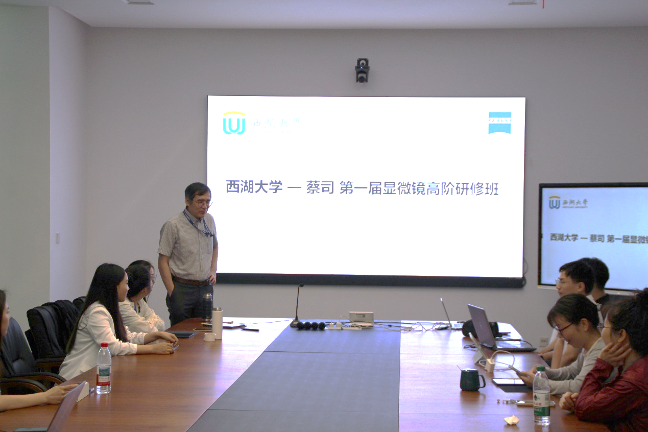
Professor Wang Yalin, Director of the Biomedical Experimental Technology Center at Xihu University, delivered a speech at the opening of the training class
Combining theory with practice, exploring the micro world together
During the 5-day advanced training course, senior engineers from Zeiss conducted systematic technical analysis on various advanced equipment such as laser confocal microscopy, fully automatic digital slide scanning system, laser layer scanning microscopy system, and lattice layer optical microscopy imaging system. The combination of "theory+practice" mode, collision of thinking, and joint exploration of the micro world with platform technicians.
Hardware engineers focus on the theoretical explanation and maintenance suggestions of the microscope's optical path principle and core optical components, and demonstrate the internal structure of the microscope system. Many on-site technicians exclaim that it is the first time they have such a clear understanding of optical path design, and express a new understanding of the equipment's principles.
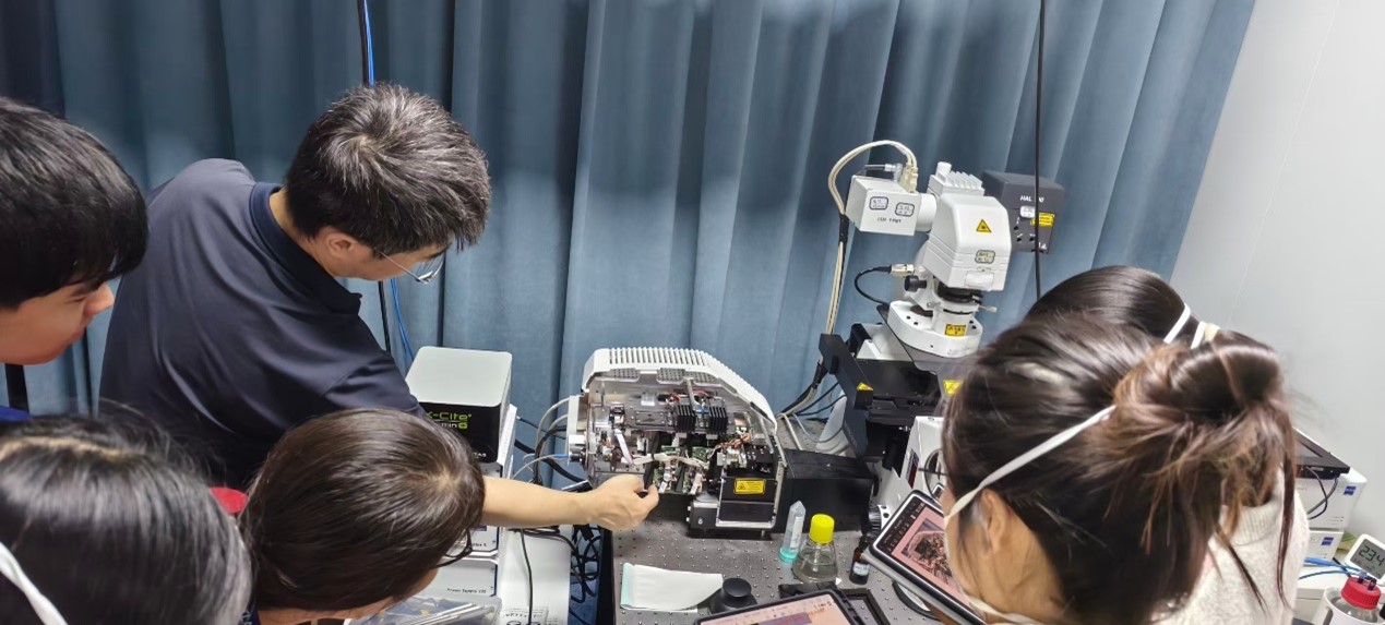
Hardware engineer showcases the internal structure of the microsystem
The application engineer, starting from the technical origin, systematically introduced the principles and evolution of imaging technology, as well as the latest imaging technologies such as instantaneous volume imaging technology Lightfield 4D and molecular dynamics tool Dynamics profiler. They demonstrated advanced functional modules one by one in the practical operation of the computer, and deeply discussed solutions to specific scientific research problems with the teachers.
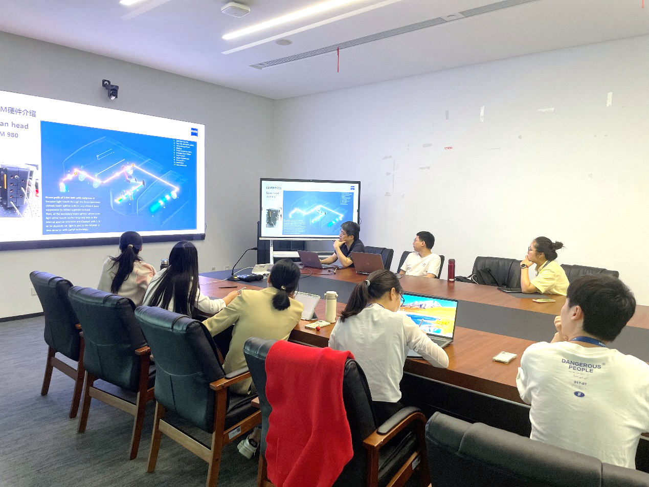
Application engineer introduces the microscope optical path

Practical training for application engineers
User oriented, empowering scientific research and innovation
As an advanced optical and optoelectronic technology enterprise, Zeiss Group has been committed to creating value for customers since its establishment in 1846. The successful holding of this training course is not only a precise docking of technology and research needs, but also opens a new chapter of deep cooperation between the two parties. In the future, we look forward to leveraging such communication platforms to better empower scientific research and innovation with advanced technology, and to facilitate the birth of more cutting-edge scientific breakthroughs.

Closing photo of advanced microscope training class