Ultra high speed imaging
Can accommodate large samples
Automated intelligent imaging process
Multi application fields
Rapid breakthrough, the essence of nanotechnology
91 parallel electron beams are imaged simultaneously, generating over 3TB of high contrast and high signal-to-noise ratio data per hour. It only takes less than 2 minutes to perform 4nm pixel size imaging on a 1mm2 slice.


Mouse brain slices. The left image uses MultiSEM 706 to image an area of 165 μ m × 143 μ m in just 1.5 seconds.
The image on the right shows the magnified effect of the 30 μ m area in a mouse brain slice. The sample was provided by B. Kasthuri from the University of Chicago.

After processing and polishing the integrated circuit, the left image was captured using MultiSEM 706 within 1.5 seconds, covering a hexagonal field of view of 132 μ m × 114 μ m. The image on the right shows the amplification effect of the 30 μ m region in an integrated circuit. The sample was provided by C. Pawlowicz from TechInsights, Ontario, Canada.
Large sample imaging, high-throughput analysis
MultiSEM enables imaging of large-area samples. Taking into account both the overall structure and details of the ultra large sample, combined with sequence slicing collection, high-resolution 3D analysis can be performed on intact cells, large volume tissues, and even intact brain tissues.

Use MultiSEM to perform high-resolution imaging of 1 mm3 human cortical tissue and perform three-dimensional reconstruction analysis of various cellular structures within it. The left image shows the three-dimensional reconstruction effect of pyramidal neurons in a 2 x 3 mm2 tissue plane. The right figure shows the three-dimensional reconstruction of local volume neurons. The image was provided by the Lichtman Laboratory at Harvard University, and the rendering was produced by D. Berger.

Reverse engineering application: Using MultiSEM to quickly complete the layered overall scanning of advanced process graphics chips with a 5nm pixel size and a 100ns point dwell time. The imaging data of the logarithmic layer structure can be 3D reconstructed to verify the layout design. It only takes 1.4 seconds to obtain nanoscale resolution images of hexagonal regions (with a long axis of 165um) through a single 91 beam simultaneous scan.
Intelligent control, simplifying complexity
The innovative ZEN software interface is intuitive and easy to use, the experimental process assistant simplifies tedious settings, multiple intelligent auxiliary functions reduce the workload of operators, and the automatic light path adjustment system ensures long-term high-quality imaging without human intervention.
Automatic slice detection, saving manual labor

Intelligent data management ensures data integrity
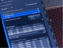
Using ZEN imaging software to control Zeiss MultiSEM
New Zeiss MultiSEM 706
Large scale ultra-thin imaging
以突破性的速度
Decrypt more mysteries