-
E-mail
sales@opton.com.cn
-
Phone
13126536208
-
Address
Oubotong Group, Building 2, No. 1100 Huihe South Street, Chaoyang District, Beijing
Product Categories
Beijing Obotong Optical Technology Co., Ltd
Thermo Fisher Scientific Field Emission Scanning Electron Microscope Standard Energy Spectrum Assisted Material Analysis
NegotiableUpdate on 01/18
- Model
- Nature of the Manufacturer
- Producers
- Product Category
- Place of Origin
Overview
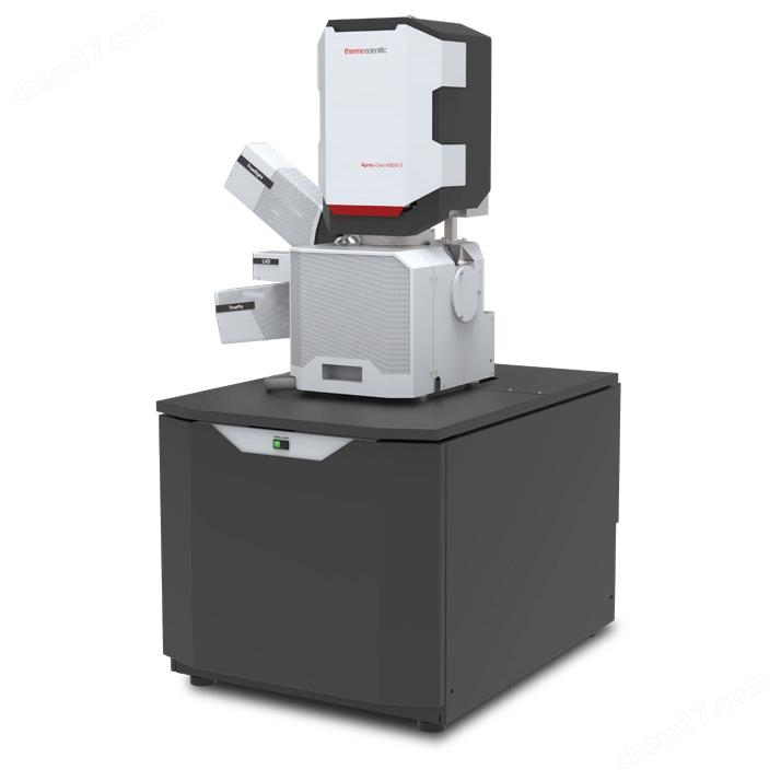
Thermo Fisher Scientific's field emission scanning electron microscope comes standard with energy spectrum assisted material analysis, Thermo Scientific Apreo ChemiSEM Field emission electron microscopy is the preferred choice for high-performance and high-quality imaging. It integrates an automated workflow for elemental composition and structural analysis, allowing users with different experiences to easily obtain high-quality data. Apreo ChemiSEM provides integrated analytical techniques and complete analysis results suitable for various samples and users with different levels of experience for large research institutions and industrial clients.
Product Details
Thermo Scientific ™ Apreo ChemiSEM ™ Field emission electron microscopy is the preferred choice for high-performance and high-quality imaging. It integrates an automated workflow for elemental composition and structural analysis, allowing users with different experiences to easily obtain high-quality data.
Apreo ChemiSEM provides large research institutions and industrial clients with solutions suitable for various samples and users with different levels of experiencewholeIntegrated analysis techniques and complete analysis results. For multifunctional laboratories with multiple users and various sample types, the system's new intelligent frame integration (SFI), autofocus, and auto astigmatism functions can automatically optimize image acquisition parameters and collect data without manual adjustment. The system always maintains a good state and can perform imaging at any time, allowing users more time to focus on obtaining the required data.

ChemiPhase is used for phase analysis of composite inclusions in steel. ChemiPhase can effectively identify various inclusions, obtain the composition and area fraction of various inclusions, and easily detect unexpected phases. Complete information can be extracted through one analysis.
Main Features
High resolution imaging performance:Thermo Scientific ™ Trinity ™ Detection and automation technology can simplify data collection in various applications
*Optimization analysis solutionsTrueSightPro EDS detector, ChemiSEM technology, and TruePix EBSD detector can fully and comprehensively characterize samples
Integrated workflowCompatible with MAPS software, custom scripts, and CleanConnect ™, Compatibility allows users to have full control over the analysis process
The Apreo ChemiSEM system integrates three features that help simplify operations and ensure reliable results: Thermo Scientific ™ ChemiSEM ™ Technology, new TruePix backscattered electron diffractometer (EBSD), and unique Thermo Scientific ™ Trinity ™ Tube detection system.

ChemiSEM technology utilizes fully automated real-time quantitative algorithms to present features that cannot be noticed solely through imaging, providing more complete information for samples. This feature runs anytime and is easy to use, providing faster results and helping to eliminate user bias.
The combination of TruePix EBSD detector and Apreo ChemiSEM system can expand its application range. The brand new EBSD software can be used to control the detector, guiding users to complete parameter settings, data acquisition, and data processing.
The Trinity tube detection system can achieve high-quality synchronous acquisition of morphology, polar surface, and composition information without the need for ETD or BSD detectors.
It has a unique signal filtering option, which can achieve a high resolution of 0.9 nm ZUI without electron beam deceleration at 1 kV voltage.
The Apreo ChemiSEM system combines high-resolution imaging, elemental analysis, and structural analysis to provide users with powerful tools for analyzing various types of materials.
electron optics
The high-resolution field emission SEM tube has the following characteristics:
- High stability Schottky field emission gun, providing stable high-resolution analysis beam current
- Composite final stage lens, combined electrostatic lens, leakage free magnetic lens, and immersion magnetic lens (optional)
-60 ° objective lens geometry, allowing for larger sample tilts
- Self heating aperture ensures cleanliness, contactless aperture switching
SmartAlign technology: No need for user alignment
In low vacuum mode (optional), the use of differential vacuum pumping through the lens reduces the scattering effect, achieving accurate analysis and high-resolution imaging of ZUI
• Electron beam deceleration function, sample stage bias range is -4000 V to+600 V
Continuous variation of beam current and optimization of pore size keratin
Dual sample stage scanning deflection
Easy installation and maintenance of electronic guns: automatic baking, automatic start-up, no need for mechanical alignment
• Pivot Beam mode, used for selective electron channel contrast imaging, also known as "Swing Electron Beam" mode (limited to Apreo ChemiSEM S-type)
ZUI short filament lifespan: 36 months
ChemiSEM technology
• TrueSight; Detector size: 25 or 70 mm ²
• Energy resolution up to 125 eV @ Mn Κα ₁
The detection range of elements is Be Am
Light element sensitivity can detect silicon (Si L α)
SEM-EDS functionality integrated into a single user interface
• Project based data storage
Project data tree, convenient and easy to manage data
Industry standard data format
• Dedicated analysis mode, achieving seamless integration between point, line, and surface scanning modes
Select the available electronic image types in the xT SEM user interface
One click report generation
• Removal of peaks and escape peaks
• Automatic peak recognition
• Combination peak and back bottom superposition
Accurate quantification over a wide range of working distances, beam currents, and electron beam energies
Users can define options to include, exclude, or quantify missing elements
Automatic or user-defined selection of KLM line system for quantitative analysis
Remove the background through digital filtering method
• Quantitative analysis without standard samples by fitting with ZUI small two product filtering method
Using PROZA matrix correction method to achieve excellent quantification of light elements
• Qualitative and quantitative line scanning, can stop testing on time or count
ChemiSEM technology, combined with electronic image algorithms to achieve rapid quantitative analysis
Quantitative surface scanning is always on, providing peak stripping for quantitative surface scanning
• Counting surface scanning, supporting unit element line selection
• Quantitative surface scanning through square kernelization (optional)
Overlay multiple scanned images onto an electronic image
User defined or automatically selected element colors
• Collect and stitch multiple fields of view through navigation montage
• Compatible with MAPS software to achieve automatic stitching of multiple fields of view
• Use points and rectangular boxes for spectrum extraction
Extract line scan data from X-ray surface scan images by flexibly selecting line direction, width, and number of points
Normalization comparison of multispectral images
Drift correction based on DCFI for surface scanning
Using chemical stoichiometry for compound analysis of borides, carbides, oxides, and nitrides
ChemiPhase conducts real-time or offline phase identification through multivariate statistical analysis
ChemiView enables offline reprocessing and report generation for different data types
TruePix EBSD detector
• Hybrid pixelated direct electron EBSD detector
A single detector consisting of a single Timepix based module
• Zero read noise, high signal-to-noise ratio
• Zero distortion
• Single particle counting
• Energy thresholding
2000 FPS frame read rate
Complete detector insertion, calibration, pattern optimization, and automatic surface scanning settings within one minute
EBSD acquisition and data processing software
Fully integrated online collection and offline data processing system
• Automatic pattern optimization: The system collects>20 EBSPs from random points in the area to be collected, achieving high-quality background calibration
ZUI high exposure measurement: The system determines the ZUI high exposure before signal saturation, and automatically integrates frames when the exposure time exceeds the ZUI high exposure
• Automatic flat field correction and contrast enhancement (with additional calibration program provided)
Automatically calibrate the center of the pattern for different detector positions, working distances, and sample tilt angles
• Can overlay any type of image
• Grain size histogram
• Noise removal and pixel enhancement
User defined template based report
Calibrate all seven crystal systems and 11 Laue groups
• Simultaneous calibration of multiple phases
Fast Fourier Transform and Pattern Contrast Quality Index
Low SEM magnification pattern center correction
EBSP's high-quality fitting kinematic simulation and superposition
Euler diagram, X/Y/Z antipolar diagram (roll, normal, transverse), Euler orientation diagram, phase diagram, and orientation difference surface scan
vacuum system
Oil free vacuum system
1 × 240 l/s turbo molecular pump
1 x PVP vortex pump
• 2 × IGP
• Sample chamber vacuum degree (high vacuum)<6.3 × 10⁻⁶ mbar (after 12 hours of pump operation)
Vacuum extraction time: ≤ 3.5 minutes
Low vacuum mode (chamber pressure of 10-500 Pa) (optional)
• Pressure limiting aperture (PLA) automatic loading device
sample stage
Standard multifunctional sample holder, which can be directly installed on the sample table and can accommodate up to 18 standard sample holders (with a diameter of 12 mm), three pre tilted sample holders, cross-sectional sample holders, and two pre tilted STEM sample carriers (optional) (38 ° and 90 °); Installing samples does not require the use of tools
Each optional STEM carrier strip can accommodate six STEM carrier nets
• Wafer and custom sample stage (optional)


Apreo ChemiSEM supports various industrial applications, including the characterization of large and heavy materials. No need to cut the sample or reduce its size, simplifying the preparation process.
System Control
64 bit GUI, equipped with Windows 10 system, keyboard, and optical mouse
24 inch LCD display screen, WUXGA 1920 × 1200 (optional second display)
• Customizable user interface
• Automatic focusing, automatic astigmatism correction, and automatic lens centering functions
Intelligent frame integration (automatic setting of acquisition parameters)
• Image registration
• Navigation montage
• Revoke and restore function
• Joystick (optional)
• Knob board manual user interface (optional)
image processor
The residence time range is 25 ns -25 ms/pixel
Up to 6144 × 4096 pixels
• File type: TIFF (8-bit, 16 bit, 24 bit), JPEG or BMP
Single frame or 4-view image display
SmartScan mode (256 frame average or integral, line integral and average, interlaced scan)
DCFI (Drift Compensation Frame Integration) mode
Digital image enhancement and denoising filters
Installation requirements
(For detailed data, please refer to the pre installation guide)
• Power supply:
- Voltage is 100-240 V AC (-6%,+10%)
- Frequency is 50/60 Hz (± 1%)
- Power consumption:<3.0 kVA (electron microscopy basic system)
Grounding resistance<0.1 0
• Environment:
- Temperature is 20 ° C (± 3 ° C)
- Relative humidity below 80%
- Stray AC magnetic field:<40 nT (asynchronous) or<100 nT (synchronous) at a line time of 20 ms (50 Hz power supply) or 17 ms (60 Hz power supply)
ZUI small door size: 0.9 m wide x 1.9 m high
Weight: 980 kg (system console)
Suggest using dry nitrogen for vacuuming
Compressed air, 4-6 bar, clean, dry, and oil-free
• System cooling device
• Noise: requires on-site investigation
Ground vibration: requires on-site investigation
• Optional active damping device
Product Parameters
Electron beam resolution

BD: Electron beam deceleration mode, WD: Working distance (unless otherwise specified, provide resolution based on high-quality working distance). By default, the system acceptance test conditions after ZUI final installation are: high vacuum 1 kV and 30 kV conditions, with immersion mode activated (if applicable).
Electron beam parameter range
Electron beam parameter range: 1 pA to 50 nA (optional 400 nA)
• Acceleration voltage range: 200 V -30 kV
• Landing energy range: 20 eV -30 keV
ZUI large horizontal field of view width: 3 mm at 10 mm WD (ZUI small magnification: 29x)
sample warehouse
• Inner width: 340 mm
• Analysis of working distance: 10 mm
• Port: 12

Characterization of magnesium oxide particles at low kV (500 V) using electron beam deceleration (BD). BD can improve morphological details and reduce charge effects.
detector
Apreo ChemiSEM can freely combine different detectors or detector zones (optional), and can simultaneously detect up to four types of signals:
Trinity detection system (inside the objective lens and inside the tube)
- T1 segmented objective lens low position detector
- T2 objective lens internal center position detector
- T3 tube high position detector (optional)
• TD: Everhart Thornley Secondary Electron Detector
DBS: Scalable partitioned pole shoe bottom backscattered electron detector (optional)
Low vacuum secondary electronic detector (optional)
DBS-GAD: Atmosphere analysis backscattered electron detector installed on the objective lens (optional)
STEM 3+Scalable Partition Detector (BF, DF, HADF, HAADF) (optional)
• Infrared CCD
• hermo Scientifc ™ Nav-Cam ™ Navigation camera (inside the sample compartment)
Similar Product Recommend


