- Phone
-
Address
3rd Floor, Building 75, No. 139 Dongyi Road, Panyu District, Guangzhou City
Product Categories
Yikangda Technology (Guangzhou) Co., Ltd

Improved Kato Katz thick smear method
NegotiableUpdate on 05/06
- Model
- Nature of the Manufacturer
- Producers
- Product Category
- Place of Origin
Overview
The Kato Katz method is a World Health Organization (WHO) method for detecting parasitic eggs, which can simultaneously detect roundworm eggs, hookworm eggs, liver fluke eggs, and more. This detection kit is used as an auxiliary tool for microscopic detection of parasite eggs in feces, which can improve the effectiveness of parasite detection and quantitatively analyze the degree of infection.
Product Details
Improved Kato Katz thick smear method
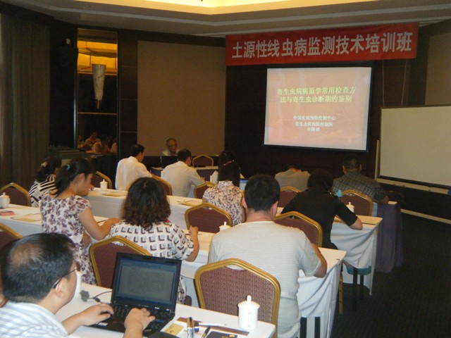
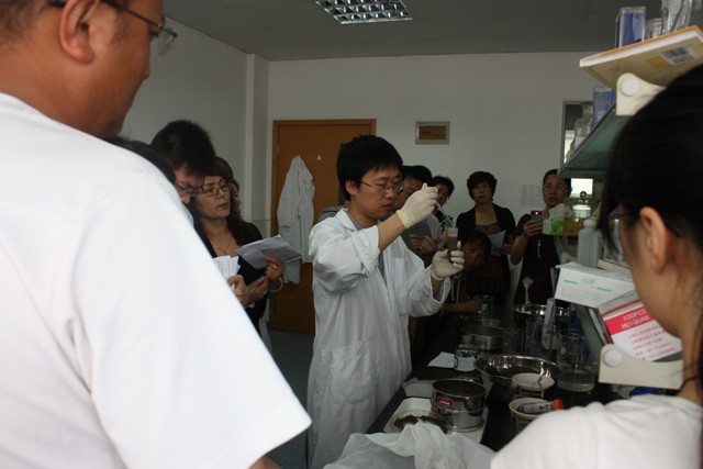
Parasitic Disease Institute holds training course on soil borne nematode disease monitoring technology |
|||||
| Host: Chinese Center for Disease Control and Prevention | |||||
In order to further promote the monitoring of soil transmitted nematode disease throughout the country and improve the technical ability of grass-roots disease control personnel in soil transmitted nematode disease prevention and control, the Institute of Parasitic Diseases held a training course on soil transmitted nematode disease monitoring in Shanghai from June 21 to 24. The training course was attended by about 20 relevant professionals from 9 provinces (districts and cities) where national monitoring points of non soil transmitted nematode disease are located, including Jilin, Liaoning, Inner Mongolia, Ningxia, Shaanxi, Tianjin, Zhejiang, Tianjin and Xizang. |
|||||
Instruction Manual for Parasite Egg Detection Preparation
(Improved Kato's method)
Introduction:The improved Kato's method is a parasitic egg detection method developed by the World Health Organization (WHO), which can simultaneously detect roundworm eggs, hookworm eggs, liver fluke eggs, and more. This detection kit is used as an auxiliary tool for microscopic detection of parasite eggs in feces, which can improve the effectiveness of parasite detection and quantitatively analyze the degree of infection.
Composition of detection package:
1. Composite dye solution 1 bottle 20ml/bottle
2. 100 quantitative plates
3. 100 scraper blades
4. 100 pieces of nylon mesh
5. 100 sheets of glass paper
6. 100 glass plates
Operation steps:
1. Place a nylon swab on the fecal sample to be tested, gently scrape it with a plastic scraper to allow fine fecal residue to pass through the holes of the nylon swab, and collect the filtered feces.
2. Place the quantitative plate in the middle of the glass slide, and then fill the fine fecal residue on the scraper into the circular hole of the quantitative plate, filling the entire hole and smoothing it out. Carefully remove the quantitative plate, leaving the fecal sample on the glass slide.
3. Take one soaked hydrophilic glass paper, shake off excess transparent liquid, cover it on the fecal sample, and gently press it with a thick glass slide to evenly spread the fecal matter under the glass paper.
4. Place the prepared Kato tablets in the specimen box (lined with absorbent paper).
5. After numbering, conduct a microscopic examination at room temperature of 25 ℃ and humidity of 75% for about 1 hour, or at a temperature of 30-36 ℃ for about half an hour.
Microscopic examination and Result judgment:
1. Microscopic examination: Place the transparent Kato film on the stage of an optical microscope and examine it under a low magnification mirror (10x). Microscopic examination carefully examines each field of view and pays special attention to the identification of unfertilized roundworm eggs. Count the entire insect eggs (counting principle: count up but not down, count left but not right).
2. The egg count of Kato tablets: The number of eggs per gram of feces (EPG) is calculated by multiplying the number of eggs detected by microscopy by 24 times (one fecal sample per test).
Calculate the number of schistosomiasis eggs in three Kato slices as A, B, and C respectively, and the number of eggs per gram of feces (EPG) is: (A+B+C) × 8 (three tests per feces).
Notes:
1Each fecal sample requires three smears (one stool and three tests to improve detection rate)
2. Cut glass paper should be twisted into pieces and placed in transparent liquid. Avoid putting it in a whole stack to prevent the inside from being soaked and affecting the transparency effect; The soaking time should not be less than 24 hours.
3. The transparency time of thin shell insect eggs, such as hookworm eggs, should not exceed 2 hours to avoid missed detection due to excessive transparency.
4. When the feces are hard, soak them in a little water or clear liquid for a moment, and then scrape them off.
5. The quantitative plate must be placed in the middle of the glass slide. If the quantitative plate is biased to one side, the fecal film at the edge of the slide cannot enter the field of microscopic inspection after slicing, resulting in missed detection.
When making specimens, excessive drying should be avoided to prevent deformation or rupture of the insect eggs, which may affect their identification.
The speed at which fecal residue becomes transparent depends on temperature, humidity, and the thickness of the fecal film. Generally, it can be left at room temperature, but in winter, it needs to be placed in a temperature chamber to accelerate transparency. If the fecal membrane is too thick, it is difficult to find the eggs.
This testing kit is stored at room temperature and is valid for 24 months.
Similar Product Recommend


