- Phone
-
Address
Beijing
Product Categories
Shilian Boyan (Beijing) Technology Co., Ltd


High throughput cell contraction force testing using porous plates and micro column arrays
NegotiableUpdate on 06/16
- Model
- Nature of the Manufacturer
- Producers
- Product Category
- Place of Origin
Overview
High throughput cell contractility testing using porous plates and micro column arrays, including high-throughput cell contractility testing using 24, 96, and 384 well plates, cell micro column arrays, and traction microscopy
Product Details
High throughput detection of cell contractility using porous plates
Background:
Cells have the ability to generate power, and they use physical force to perform important functions - when this power is disrupted, it shortens and prolongs while performing important functions, leading to disease. Traditionally, measuring such forces was difficult, but the following two new methods can now simultaneously measure the forces produced by thousands of cells.
Test plan:
1、 High throughput detection of cell contractilityPorous plate-24, 96, 384 well plates:
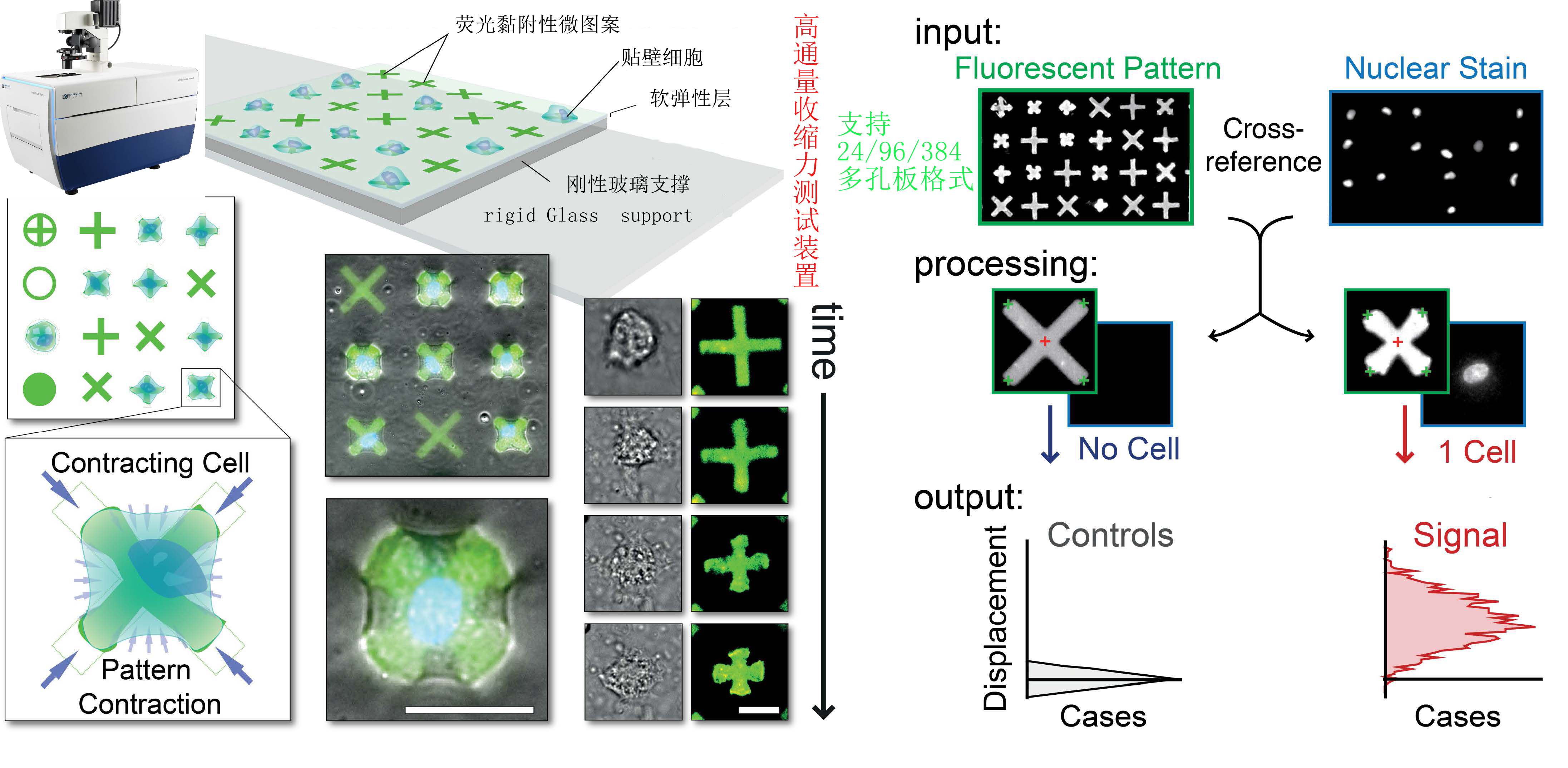
Figure 1: Schematic diagram:
1.1) Cell Contraction Force Test 24 well Plate
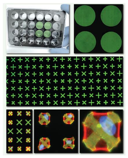
1.2)Cell Contraction Test 96 well Plate
1.3) Cell Contraction Force Test 384 well Plate
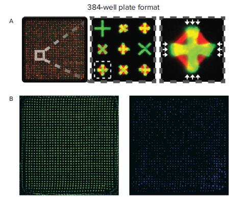
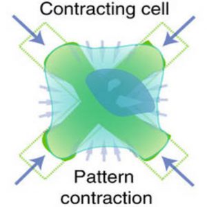
The porous plate capable of high-throughput detection of cell contraction force isA microporous plate with fluorescent micro patterns embedded in the bottom of the soft film,There are over 100000 evenly distributed X or+shaped adhesive protein micropatterns on top, on which cells settle and attach,The X embedded in the board has elasticity,Therefore, whenever cells contract, they will contract.X carries molecular markers that can emit fluorescence, allowing for imaging and quantification of reduced shapes.
It can be imaged and quantified, so researchers can associate the deformation of X or cross with a certain amount of force on a single cell or thousands of cells. This technology also provides researchers with the ability to examine individual cell calcium responses and their relationship with cell strength.
This technology can also be used for drug discovery. Researchers can quickly and easily test different therapeutic molecules to understand how they affect cellular forces and whether any potential force issues can be corrected.
Having controllable rigidityThe elastic film is embedded with user designed adhesive and fluorescent micro patterns, forming a very dense but uniform array (>120 microns/square millimeter). 100000 patient derived single cells (obtained from human tissue at the origin of the disease) are independently located and adhered to micro patterns (one cell per micro pattern), exerting traction on the micro pattern and significantly changing its shape, enabling intuitive image-based evaluation of cell contractility at a throughput of *.
1) Micro patterning of Fluorescent Elastic Shrinkable Surface
- With controllable rigidityThe elastic film is embedded with user designed adhesive and fluorescent micro patterns, forming a very dense but uniform array (>120 microns/square millimeter). 100000 patient derived single cells (obtained from human tissue at the origin of the disease) are independently located and adhered to micro patterns (one cell per micro pattern), exerting traction on the micro pattern and significantly changing its shape, enabling intuitive image-based evaluation of cell contractility at a throughput of *.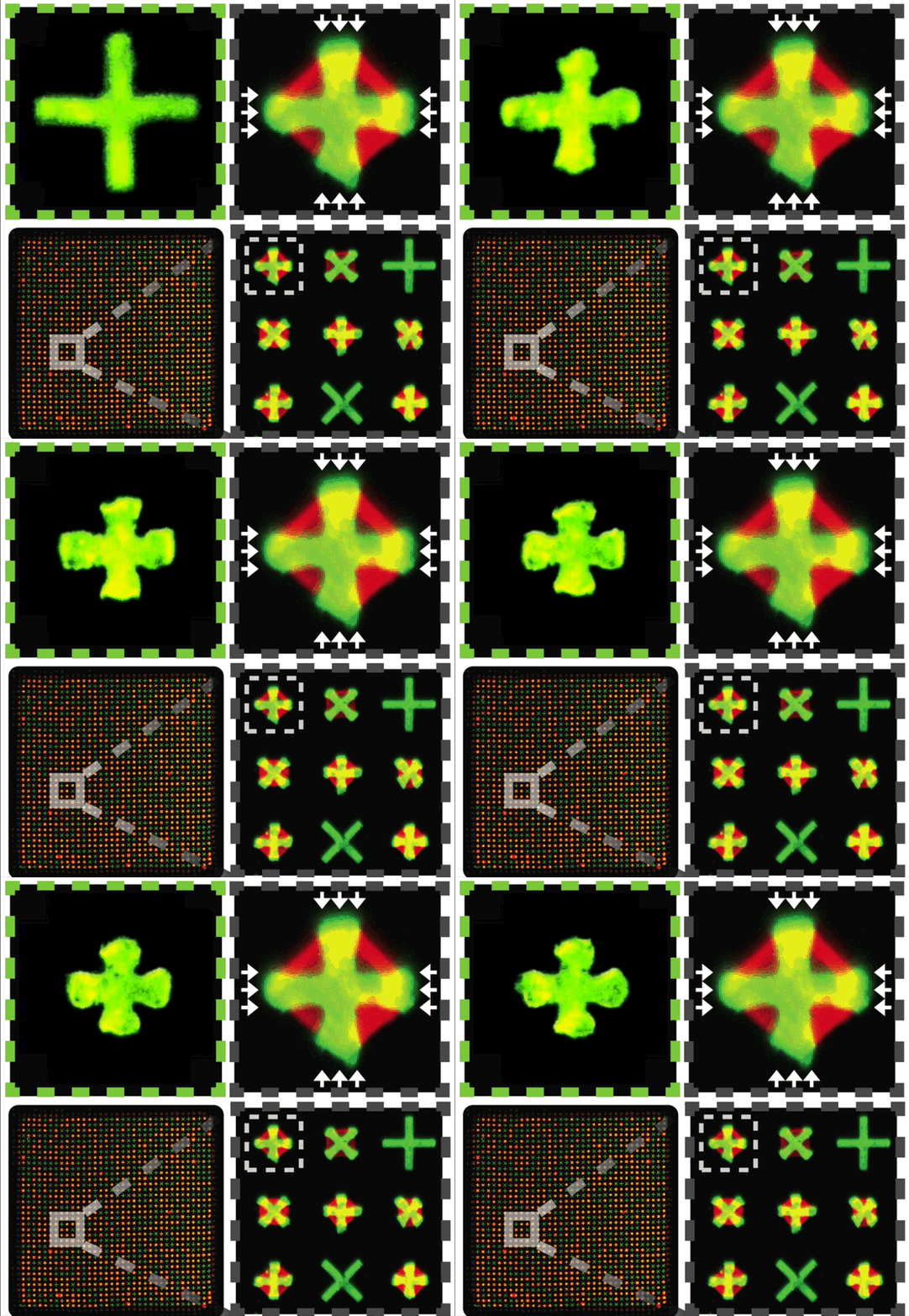
2) Dynamics visualization of controlled single-cell contractility based on images
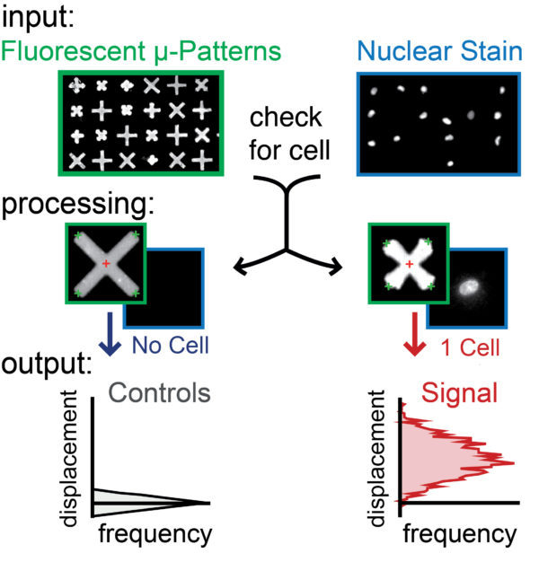
For example, the micro patterns of each attached cell in a 384 well plate are independently monitored with fine temporal resolution over extended periods of time to directly observe the full range of contraction behavior, from tension induced contraction to induced contraction or relaxation, as well as the window of action for any effect. Select the "x" - shaped micro pattern to reduce the cell substrate contact area and increase the cell diffusion area, thereby generating amplified and concentrated forces at different positions. Due to the mechanical separation of each micropattern from other micropatterns, adhered cells do not transmit strain to adjacent micropatterns, ensuring reliable evaluation of the contractile force of each individual cell in the population.
3) Automated and intuitive image analysis of single-cell contractility
Based on this method, detection generates intuitive and clear contraction signals - providing direct image analysis to obtain quantitative and reliable data from each single cell in the imaging population.
Can simultaneously obtain contractility data of over 1000 uniformly patterned single cells and seamlessly integrate with 96- and 384 well plate formats to facilitate large-scale drug screening
2、 Introduction to Cell Traction Microscopy and Micro Column Array
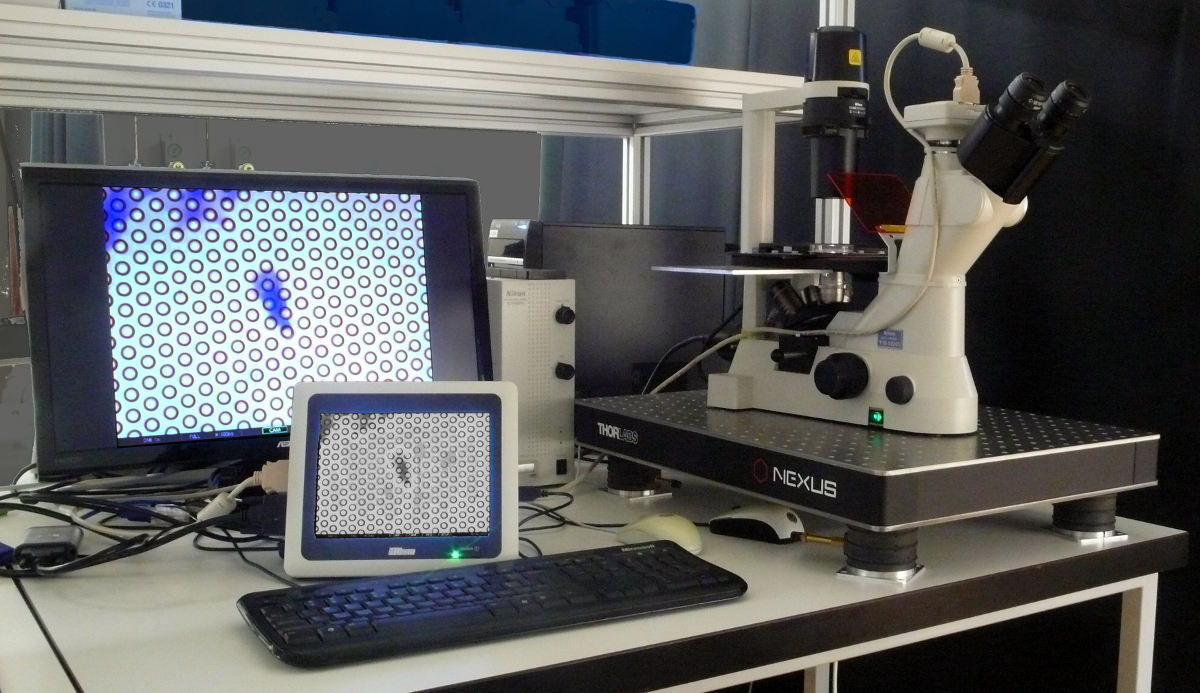
Figure 4: Cell Traction Microscopy
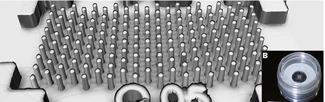
Figure 5: Cell micro column array,Customizable stiffness, size, shape, and spacing.
Shilian Boyan specializes in the sales of equipment for cell mechanics and microstructure material mechanics, providing equipment such as cell stress loading culture and micro mechanics testing and analysis systems. Click here to view more
Similar Product Recommend


