-
E-mail
info@boyuesh.com
-
Phone
15921886097
-
Address
3A, Building 28, Songjiang High tech Park, Caohejing Development Zone, Lane 518, Shenzhuan Road, Songjiang District, Shanghai
Product Categories
Boyue Instrument (Shanghai) Co., Ltd


Bruker nanoscale 3D X-ray microscope
NegotiableUpdate on 02/11
- Model
- Nature of the Manufacturer
- Producers
- Product Category
- Place of Origin
Overview
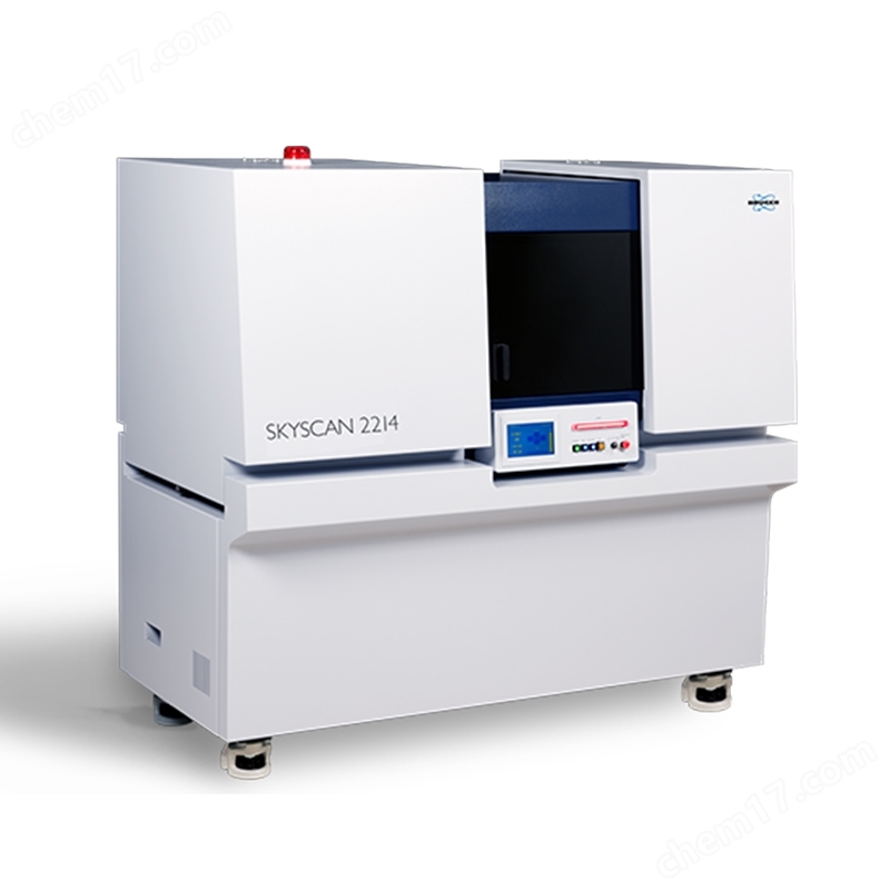
The Brooke BRUKER nanoscale 3D X-ray microscope SKYSCAN 2214 (CMOS) is a multi dose X-ray nanoscale tomography system equipped with a new generation of CMOS detectors for superior performance
Product Details
Brooke BrukerNano scale 3D X-ray microscope
SKYSCAN 2214 CMOS

SKYSCAN 2214 (CMOS) is a multi dose X-ray nanoscale tomography system. The 16 Mp large-sized sCMOS detector provides excellent resolution and expands a huge 3D field of view. Quickly switch between tungsten filaments with high penetration of 160 keV and lanthanum hexaboride filaments with excellent resolution; A true 3D spatial resolution of<500 nm can resolve two features in the generated 3D model.
thisBruker nanoscale 3D X-ray microscopeOne instrument can cover a wide range of object sizes and spatial resolutions. It has opened up new possibilities for three-dimensional imaging and precise modeling of geological materials, suitable for preclinical applications in vitro such as oil and gas exploration, composite materials, lithium batteries, fuel cells, electronic components, as well as lung imaging or tumor angiogenesis. For large objects with a diameter of over 300mm, this instrument can scan and reconstruct their internal microstructure in 3D without damage; For small-sized samples, this instrument can achieve sub micron resolution.
SKYSCAN 2214 is equipped with 3D.SUITE software. This comprehensive software includes GPU accelerated reconstruction, 2D/3D morphology analysis, and surface and volume rendering visualization.

SKYSCAN 2214light source
SKYSCAN 2214 adopts a new generation of open X-ray source. This light source can achieve actual spatial resolution better than 500 nm, X-ray energy up to 160 keV, and power up to 16 W. Due to its simple pre registered filament replacement program, the light source requires almost no maintenance.
SKYSCAN 2214 features an open (pumped) nano focal X-ray source with a diamond window. It can generate X-ray beams with peak energies ranging from 20 kV to 160 keV and provides two types of cathodes. Tungsten (W) cathodes are suitable for a complete acceleration voltage range of up to 160 kV, with a spot size of up to 800 nm. Lanthanum hexaboride (LaB6) cathodes are suitable for acceleration voltages from 20 kV to 100 kV, and the spot size of the X-ray beam can be less than 500 nm, ensuring excellent resolution in imaging and 3D reconstruction. The JIMA resolution test card shows that it can easily resolve structures up to 500 nm.
In order to ensure the long-term stability of the focal spot size and the position of the emission source, the X-ray source can also be equipped with a water cooling system, which includes a circulation device that can accurately control the temperature of the cooling liquid to maintain temperature stability.

SKYSCAN 2214detector
To achieve excellent flexibility, the SKYSCAN 2214 can be equipped with four X-ray detectors: three CMOS cameras with different resolutions and fields of view, as well as a large-sized flat panel detector. All cameras can be selected by clicking the mouse. Different CMOS cameras can be modified at any time during the system lifecycle.
All three CMOS cameras are capable of capturing images at the center of the beam and two offset positions, thereby doubling the field of view. Through offset compensation and intensity difference correction, images taken at two offset positions can be automatically stitched together.
When using small pixel CMOS detectors, high-resolution imaging and 3D reconstruction can also be performed on large-sized objects. The flexibility of the built-in detector allows it to adjust the field of view and spatial resolution according to the size and density of the object. Through excellent local reconstruction of large samples, it can scan specific components of a large-sized object with high resolution and obtain equally high-quality images.
In addition, by utilizing the offset of the detector and the vertical movement of the object, the field of view can be expanded in both horizontal and vertical directions.

SKYSCAN 2214sample room
SKYSCAN 2214 has a highly precise sample stage that supports objects with diameters up to 300 mm and weights up to 20 kg. The air suspension rotary motor can accurately rotate the position of objects with very high accuracy, and the integrated precision positioning platform can ensure sample alignment.
A large and user-friendly sample room for scanning large objects and installing optional test benches. It has enough space to accommodate peripheral devices.

SKYSCAN 2214In situ test bench
Brooke's material testing platform is capable of conducting compression tests at 4400 N and tensile tests at 440 N. All test benches can be automatically connected through the system's rotating table without the need for any external cables. By using the provided software, a scheduled scanning test can be set up.
Brooke's heating and cooling stations can reach temperatures of+80 º C or 30 º C lower than the ambient temperature. Like other test benches, the heating and cooling benches do not require any additional connections, and the system can automatically identify different test benches. By using heating and cooling tables, samples can be tested under non environmental conditions to evaluate the effect of temperature on the microstructure of the samples.
SKYSCAN 2214 is compatible with the DEBEN test bench. With the built-in adapter, the DEBEN test bench can be easily installed on the rotating table of SKYSCAN 2214.

SKYSCAN 2214Location, scanning, reconstruction, and analysis
The Brook XRM solution includes all the software required for collecting and analyzing data. Intuitive graphical user interface, user guided parameter optimization support for experts and novice users. By using a new GPU powered algorithm, the reconstruction time is greatly reduced. CTVOX, CTAN, and CTVOL are combined into a powerful software suite for qualitative and quantitative analysis of models.
·Measurement software:
SKYSCAN 2214- Instrument Control, Measurement Planning, and Collection
·Rebuilding software:
NRECON - Convert 2D Projection Images to 3D Volumes
·Analysis software:
DATAVIEWER - Slice by slice inspection for registration of 3D volumes and 2D/3D images
CTVOX - Realistic Visualization through Volume Rendering
CTAN-2D/3D Image Analysis and Processing
CTVOL - To export surface model visualization for CAD or 3D printing


SKYSCAN 2214Application Direction
- Mineralization organization
SKYSCAN 2214 can provide nano CT imaging performance, equipped with four CMOS cameras and a large-sized 6 megapixel cMOS tablet for imaging high voltage and large bone or dental samples; Three small format cameras are used to provide resolution and X-ray energy windows for scanning each bone sample.
Submicron voxel scanning can provide crystal clear bone cell fractures and microscale mineralized structures, as well as nanoscale resolution of biomaterial scaffold structures.
Phase retrieval (Paganin) provides a new dimension for analyzing bone micro mineralization patterns that have been rarely studied to date.
Orthopedic research using sheep, primates, or similar models can be conducted using a 14 cm scanning field of view and a 160kV X-ray source. Achieve your orthopedic and biomechanical research goals through mechanical testing and temperature control phases.
Morphological measurement has comprehensive 3D and 2D parameters, while density measurement includes BMD calibration reference within the preclinical size range. Excellent 3D image analysis functions include 3D registration, adaptive thresholding, Euler connectivity, fractal, anisotropy and stereo, filtering, Boolean logic operators, etc.

- soft tissue
Supporting in vitro nano CT scanning of biological tissues, with pixel sizes of sub microns, similar to histology or electron microscopy, but in true, deep 3D - this is a wonderful method for non-destructive display of internal structures. Contrast agents or chemical drying can improve image quality by further enhancing or distinguishing tissue density. SKYSCAN 2214 plays a leading role in the emergence of new imaging disciplines - micro CT histology and tissue morphometry. The multifunctionality of the system ensures that each sample can be scanned with optimized parameters, excellent resolution, and contrast.
The device is equipped with four CMOS cameras and one large flat panel camera with a resolution of 6 million pixels, which can be used for imaging high voltage and large tissue samples; Three small format cameras provide resolution and X-ray energy window for sample scanning. Comprehensive 3D image analysis capabilities, including morphological and density measurements, 3D registration, segmentation, and image processing methods.

- Plants and animals
Micro CT provides better visualization of the subtle details of internal biological structures. This imaging method can create a nanoscale 3D X-ray attenuation map without harming or damaging the scanned object. Almost all biological tissues can be visualized and analyzed with little or no special sample processing required. Sub micron voxel resolution enables imaging of small insects, plants, or seed structures with powerful magnification and rich details.
The space in the scanning room and the precision of the rotating table ensure that all types of samples can be scanned, from small zebrafish and preserved zoological and botanical samples to potted and rock buried fossils.
Four CMOS cameras can be configured, among which a large flat panel camera with 6 million pixels can be used for imaging high voltage and large samples; Three small format cameras provide resolution and X-ray energy windows for scanning biological samples. The software suite can perform morphological measurements using comprehensive 3D and 2D parameters, and excellent 3D image analysis functions include 3D registration, adaptive thresholding, Euler connectivity, fractal, anisotropy and stereo, filtering, Boolean logic operators, etc.

Similar Product Recommend