-
E-mail
info@phenom-china.com
-
Phone
18516656178
-
Address
Room T5705, Shanghai Hongqiao Libao Plaza, No. 88 Shenbin Road, Hongqiao Town, Minhang District, Shanghai
Funa Scientific Instruments (Shanghai) Co., Ltd
Detailed Explanation of Scanning Transmission Electron Microscopy | Application and Advantages of SEM-STEM Technology in Biology
Date: 2025-10-24Read: 2
Scanning transmission electron microscope
Scanning electron microscopy (SEM) has long provided us with high-resolution three-dimensional morphology of cell and tissue surfaces. However, the mystery of life goes far beyond the surface, revealing the internal ultrastructure of cells, bacteria, and even viruses is the key to understanding life activities and disease mechanisms. Transmission electron microscopy (TEM) has always been a tool for observing and studying ultrastructure, but its expensive price and complex operation make it mostly equipped by top teams or large electron microscopy centers.
With the increasing demand for molecular localization and structural details within cells in life science research, SEM manufacturers have begun to combine scanning transmission electron microscopy (STEM) technology with SEM, successfully integrating the "perspective" capability into more popular scanning electron microscopy platforms, enabling researchers to use this internal structure analysis tool. In life science applications, SEM-STEM cleverly builds a practical bridge between optical microscopes (~200 nm) and traditional transmission electron microscopes (<0.2 nm). The resolution of modern field emission SEM-STEM can reach below 1 nm, which is sufficient to clearly distinguish most organelles, viruses, and biomolecule complexes.
SEM-STEMCore advantages brought to biological research
Compared with traditional transmission electron microscopy, SEM-STEM technology has shown significant advantages in convenience, friendliness to biological samples, and overall cost-effectiveness, making it a more universal high-resolution imaging tool in biological laboratories.
01. Easy to operate
One of the core advantages of SEM-STEM is its highly simplified operational process. Modern scanning electron microscopes have been optimized for a long time, making operation simpler (especially desktop scanning electron microscopes), and most parameter adjustments are automatically completed. This greatly reduces the threshold for device usage, allowing researchers to independently complete high-quality imaging without the need for a deep electronic optical background. In contrast, the operation of traditional TEM is still very complex and often requires the support of dedicated technical personnel. In addition, the workflow efficiency of SEM-STEM is higher. The entire process from placing the sample to obtaining high-definition STEM images usually takes only a few minutes and supports rapid vacuuming and real-time preview. This makes it particularly suitable for rapid screening and preliminary diagnosis of large samples, greatly improving research efficiency.
02. Low voltage imaging
SEM-STEM typically uses lower acceleration voltages (such as 5-30 kV), while traditional TEM often uses high voltages above 80 kV to achieve high resolution and penetration. This difference is crucial for radiation sensitive biological samples.
Low acceleration voltage reduces electron beam damage to sensitive biological samples, better preserving the original morphology and biological state of the samples. Meanwhile, low-voltage imaging can enhance the scattering signal of light element materials in the sample, thereby naturally improving the image contrast. This means that even for unstained or lightly stained biological samples, SEM-STEM is more likely to obtain images with good signal-to-noise ratio and clear structure, simplifying the sample preparation process.
03. High cost-effectiveness
In terms of purchase and maintenance costs, a scanning electron microscope equipped with STEM function is much lower than a traditional transmission electron microscope. For most biological laboratories, their research questions do not always require atomic resolution, so a SEM-STEM that can provide excellent surface morphology and sufficient internal structural information is a more cost-effective choice.
Pharos STEM: tailor-made imaging solutions for life sciences
Pharos STEM desktop field emission bioelectromicroscope is a powerful desktop scanning electron microscope designed with integrated STEM capabilities specifically for life science research. By using Schottky field emission electron guns, laboratory grade high resolution is achieved within a compact desktop body, bringing high-performance microscopy imaging capabilities directly into your daily research environment.

01. Break free from the shackles of space
Pharos STEM adopts a desktop design, with a compact size that only requires a standard laboratory table to complete installation. Its built-in seismic system effectively isolates environmental interference, ensuring image stability even in high-rise buildings or vibration sensitive experimental environments.
02. Simplicity
Pharos STEM simplifies complex operations into an intuitive process: injecting samples while vacuuming → switching to STEM mode with one click → automatic focusing, adjusting brightness and contrast → clicking to capture high-definition images. 15 second ultra fast vacuum pumping, 40 second imaging, no need for professional operation, can be independently operated within 1 hour of training, suitable for teaching and routine research.
03. High resolution
STEM resolution better than 1.0 nm, meeting the research needs in the field of life sciences. Whether observing the internal structure of biological samples, the distribution of viruses within cells, or the state of nanoscale drug carriers after being engulfed by cells, clear and reliable results can be provided.
04. Friendly sample preparation process
Pharos STEM is compatible with standard TEM carrier networks, allowing users to directly place biological samples cut by ultra-thin sectioning machines or carrier networks containing virus or protein samples into electron microscopes for analysis, seamlessly integrating with the existing sample preparation process in biological laboratories.
Pharos STEM Application Case
01 Pathological section
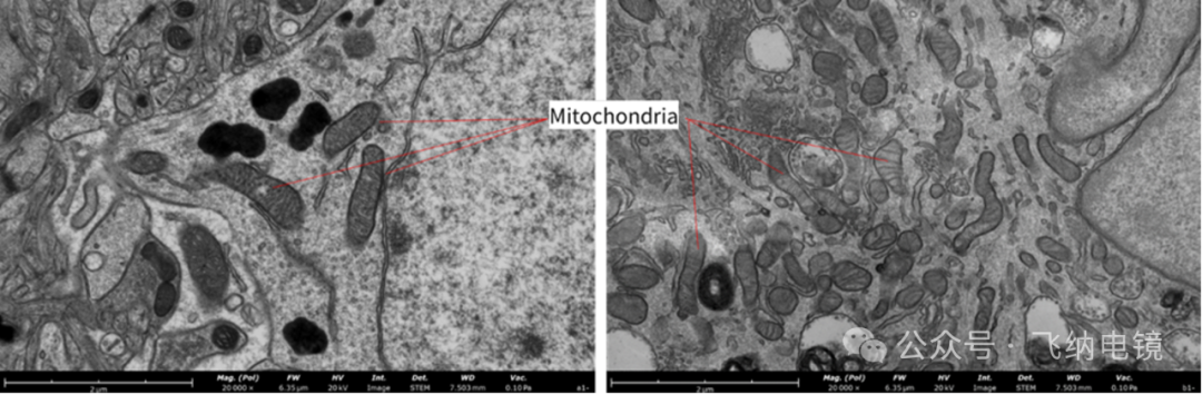
Membranous nephropathy is a common glomerular disease and one of the more common types of nephrotic syndrome. The main lesion site is the filtration membrane of the glomerulus, and pathological diagnosis is made by observing the deposition of electron dense substances. The glomerular filtration membrane structure can be seen under Pharos STEM.

02 Low voltage imaging
Due to the high acceleration voltage (80-120 kV) of transmission electron microscopy, its ability to penetrate biological samples is too strong, resulting in low contrast of transmission images. In contrast, the acceleration voltage of Pharos STEM is lower (0-20 kV), so when observing such samples in STEM mode, it can significantly improve the contrast of the transmission image. It can be used for the observation of small-sized samples such as exosomes, viruses (AAV, tobacco mosaic virus), protein particles, plasmids, etc.
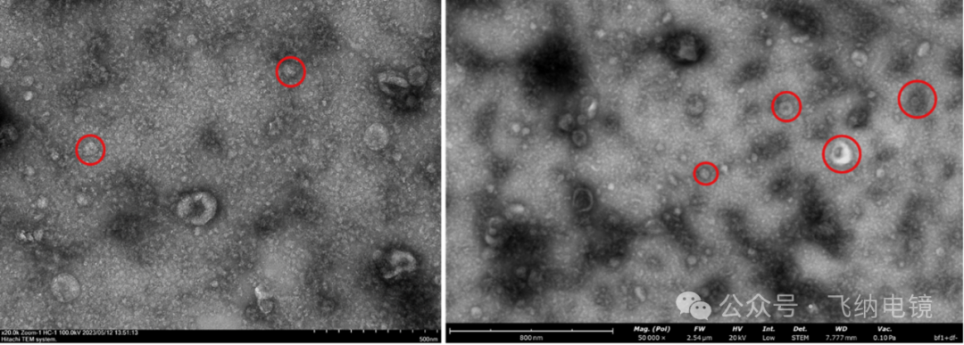
Left: Extracellular vesicles (TEM); Right: Exosomes (Pharos STEM)
03 unstained sample
When observing biological samples using transmission electron microscopy, there is often a problem of insufficient contrast. In this case, heavy metal staining such as uranium and lead is needed to obtain structural information, but the staining process is cumbersome and may alter the original structure of the sample. In contrast, unstained samples under Pharos STEM are also clearly visible, simplifying your sample preparation process and reducing sample shrinkage and heavy metal precipitation artifacts caused by staining.