-
E-mail
info@phenom-china.com
-
Phone
18516656178
-
Address
Room T5705, Shanghai Hongqiao Libao Plaza, No. 88 Shenbin Road, Hongqiao Town, Minhang District, Shanghai
Funa Scientific Instruments (Shanghai) Co., Ltd
What is Microscopic CT? What is the difference between X-ray, scanning electron microscopy, and ultrasound testing? Understand once
Date: 2025-10-24Read: 2
In daily life, CT scans that we are familiar with mostly appear in hospitals, where doctors use them to assess the internal health status of the body. But you know what? This technology has long been "out of hospitals" and entered the fields of scientific research and industrial testing, with resolution increased to the micrometer or even sub micrometer level, revealing the internal mysteries of various materials, life sciences, and cultural relics. This is micro CT.

In this article, we will take you through a comprehensive understanding of micro CT, how it differs from X-ray equipment, scanning electron microscopy, and ultrasound testing, and in which fields it can play a role.
PART.01 Birth and Development of Microscopic CT
In 1895, German physicist Wilhelm Conrad R ö ntgen accidentally discovered a new type of radiation that could penetrate the human body and objects while studying cathode rays. He used his wife's hand to make the first X-ray, thus opening the prelude to the "perspective era" of medicine and material testing. R ö ntgen also won the first Nobel Prize in Physics for this.
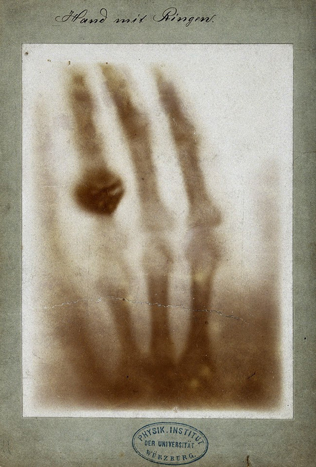
X-ray photograph of Mrs. R ö ntgen's hand, taken on December 22, 1895
Over the next century, X-ray imaging continued to develop, from early planar projection to the later emergence of computed tomography (CT), achieving three-dimensional reconstruction of the human body and the interior of objects. Traditional CT is mostly used for medical diagnosis, while in scientific research and industrial testing, people require higher resolution and more precise structural observation, so "Micro CT" has emerged.
PART.02The imaging principle of micro CT
Microscopic CT technology uses X-rays to irradiate samples, records the intensity distribution of transmitted X-rays through detectors, and then reconstructs the three-dimensional internal structure of the sample using computer algorithms. Its uniqueness lies in its ability to provide high-resolution and three-dimensional images in a non-destructive manner.

Microscopic CT technology can provide detailed three-dimensional internal information of materials without damage, including:
1Structural information:Such as diameter, volume, surface area, roundness, connectivity, spatial distribution .....
2Density information:Such as cavity pores, element weight, and composition distribution .....
33D model:Such as finite element analysis and 3D printing .....
Application of PART.03 Microscopic CT
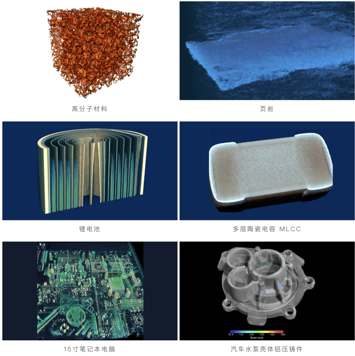
Microscopic CT technology has become a tool for scientific research and industrial applications due to its non-destructive nature, ultra-high resolution, and three-dimensional imaging capabilities. At present, it has been widely used in industries such as orthopedics, agriculture, archaeology, 3D printing, pharmaceuticals, materials science, geology, food science, electronic semiconductors, lithium batteries, automobile manufacturing, aerospace, etc. The following results were observed and imaged using Neoscan high-resolution micro CT.

PART.04 Differences between Microscopic CT and X-Ray
Many people confuse micro CT with common X-ray equipment, but in fact, although both use X-ray principles, the imaging methods and application scenarios are very different.

The difference between X-ray and micro CT (image source network)
Different imaging methods:
X-Ray equipment: Obtain a two-dimensional projection image through a single exposure, similar to a black and white photograph, with different density areas inside the object overlaid on the image.
Microscopic CT: The sample rotates between the X-ray source and detector, and the system collects hundreds or thousands of projection images. After computer tomography reconstruction, the three-dimensional structure of the sample is generated.
Different dimensions of information:
X-Ray equipment: can only see "overlaid shadows", difficult to distinguish between front and back layers.
Microscopic CT: It can observe layer by layer like a "slice" to obtain a clear internal 3D model.
Resolution and applicable range are different:
X-Ray equipment: With fast imaging speed and wide range, it is suitable for quickly detecting large defects in industrial non-destructive testing, such as whether solder joints are hollow or whether parts have cracks.
Microscopic CT: With higher resolution, up to micrometer or even sub micrometer level, it can analyze micro pores, fibers, and tissue details inside materials, making it more suitable for scientific research, precision manufacturing, and structural analysis.
PART.05 Differences between Microscopic CT and Scanning Electron Microscopy
Microscopic CT and scanning electron microscopy are both important tools for microscopic research, but they focus on different directions.
Scanning electron microscopy uses an electron beam to scan the surface of a sample with a very high resolution, up to the nanometer level, and can present clear surface morphology and grain details. But it can only see the surface layer and cannot reveal internal information, and the sample usually requires conductive treatment or gold spraying.
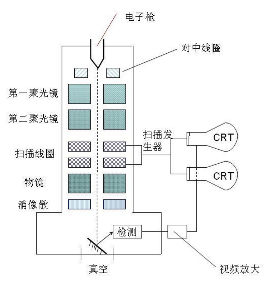
Schematic diagram of scanning electron microscope imaging
Microscopic CT relies on X-ray penetration imaging to display the three-dimensional internal structure of the sample, making it particularly suitable for studying the pore network, crack propagation, and multiphase distribution of materials. Although its resolution is lower than that of electron microscopy, it can see the "depths" that electron microscopy cannot reach.
The two are often used in combination: first, internal defects are discovered using micro CT, and then details are analyzed by slicing and electron microscopy.

Using Neoscan micro CT to locate the through-hole position of IC samples first
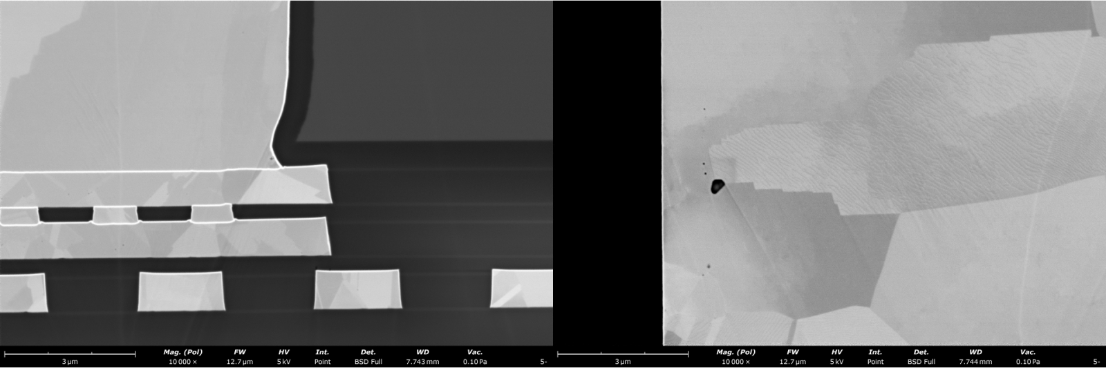
After ion milling treatment, the sample was placed in a Funa electron microscope to observe details
PART.06 Differences between Microscopic CT and Ultrasonic Testing
Ultrasonic testing relies on the reflection of sound waves during propagation in materials to determine the location of defects, and is commonly used for non-destructive testing of large components. Its advantage is that it has a large penetration depth and can detect cracks in thick walled materials, but its resolution is low and the results rely more on signal interpretation, making it difficult to visually display defect morphology.
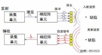
Schematic diagram of ultrasound detection imaging (image sourced from the internet)
Microscopic CT uses X-rays instead of sound waves, with higher resolution and clear three-dimensional imaging results. It is particularly suitable for the fine structure research of small and medium-sized samples, which can intuitively see the crack direction, pore distribution, and material interface.
PART.07 Summary
Therefore, micro CT can be used in conjunction with other detection methods to form complementary advantages. Through this combination of multiple technologies, researchers and engineers can obtain comprehensive sample information at different scales: micro CT non-destructive 3D internal structure imaging, electron microscopy supplementing high-resolution surface details, X-ray and ultrasound detection achieving rapid pre screening and overall evaluation, thereby achieving more accurate and efficient detection and analysis.