-
E-mail
bder1949@126.com
-
Phone
17811984612
-
Address
Room 1626-1628, Exhibition Center, No. 2288 Zuchongzhi Road, Pudong New Area, Shanghai
Product Categories
Bozhuo Biotechnology (Shanghai) Co., Ltd


Preclinical animal ultrasound microbubble contrast agent
NegotiableUpdate on 01/20
- Model
- Nature of the Manufacturer
- Producers
- Product Category
- Place of Origin
Overview
USphereTM bubble contrast agent is a preclinical animal ultrasound microbubble contrast agent (for scientific research purposes only). After intravenous injection, it can enhance the ultrasound signal in the blood, achieving the purpose of enhancing cardiovascular disease diagnosis or tumor detection. This ultrasound contrast agent uses decafluorobutane (C4F10) gas as the core gas and a single-layer membrane microbubble with phospholipids material as the shell layer.
Product Details
USphereTM Ultrasound Microbubble Contrast Agent Product Introduction
1、 Product Introduction
1.1 Physical Structure
USphereTM bubble contrast agent 系Preclinical animal ultrasound microbubble contrast agent(For scientific research purposes only), after intravenous injection, it can enhance the ultrasound signal in the blood, achieving the purpose of enhancing cardiovascular disease diagnosis or tumor detection. This ultrasound contrast agent uses Octafluoropropane (Perflutren, C3F8) gas as the core gas and a single-layer membrane microbubble with phospholipids material as the shell layer. As shown in Figure 1, through the self-assembly ability of phospholipids, the phospholipid layer can form an effective protective film to slow down gas diffusion, prevent microbubbles from melting into larger bubbles, and maintain stable circulation in the body, enhancing contrast time. The phospholipid shell is usually composed of 2-3 types of phospholipids; The surface is modified with polyethylene glycol (PET) to avoid aggregation between phospholipid shells, and its biocompatibility is also improved.
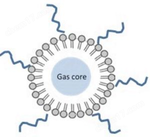
Figure 1 Schematic diagram of microbubble structure
Figure 1 Structure of Microbubbles
1.2 Shell Composition
At present, the mainstream shell materials for ultrasound contrast agents are mainly phospholipids, which have the advantage of forming a good elastic single-layer membrane to protect microbubbles and generate stable vibrations during ultrasound emission, thereby exhibiting excellent acoustic properties and enhancing ultrasound imaging effects.
1.3 Gas composition analysis
Currently, most commercially available ultrasound contrast agents use perfluorocarbons. Compared to air, perfluorocarbons have a larger molecular weight, extremely low water solubility, and slow diffusion rate; Therefore, the theoretical energy barrier for perfluorocarbon compounds to penetrate phospholipid membranes is relatively high. Perfluorocarbons can effectively slow down the deflation rate of microbubbles, thereby enhancing the contrast time of microbubbles. The gas composition used by USphereTM is also perfluorocarbon.
2、 Comparison and Advantage Analysis of Competitive Products
Common commercial ultrasound contrast agents include OptisonTM (GE Healthcare) from the US and Definity ® (Lantheus Medical Imaging)、 European SonoVue TM (Braco) and Japanese Sonazoid TM (GE Healthcare). The comparison table of common commercial contrast agents is shown in Table 1.
Advantage 1: UsphereTM has a very excellent particle size distribution compared to other products (as shown in Figure 2), and its microbubble resonance frequency is more suitable for the current range of medical instruments, greatly improving the intensity of ultrasound signals. Secondly, a good particle size distribution can control the biological effects caused by stable cavitation and inertial cavitation, thereby reducing their potential risk in detecting cardiovascular diseases and enhancing their safety in drug delivery.
Advantage 2: The UspherTM series products have smaller particle sizes, providing more detailed imaging quality while still maintaining backscatter intensity in deep tissues.
Advantage 3: The concentration of microbubbles per unit volume of UspherTM can reach 2.5x10¹⁰Bubbles/mL, which is the highest concentration among similar products. Due to its small particle size and high concentration, angiography of the tumor site is more pronounced.
Advantage 4: UsphereTM has good stability and suspension characteristics (as shown in Figure 3), and can be used for certain acoustic studies (such as ultrasonic field scanning and fluid state analysis).
Advantage 5: The circulation time of UspherTM in vivo can reach 6-15 minutes (as shown in Figure 4), which is beneficial for in vivo experiments. After opening and activating, it can maintain high quality for three days, making it convenient for in vivo experiments. However, SonoVue TM, which currently has a high sales volume, only has a 6-hour usage time after opening.


Figure 2 Physical characterization of SphereTM: Left:Electron microscopy image (Cryo TEM); Right:Particle size distribution map (Multisizer3, USPcompliant)
Figure 2. Characterization of USphereTM. Left:TEM (Cryo-TEM); Right: size distribution (Multisizer3,USP compliant)
Table 1 Common Commercial Contrast Enhanced Ultrasound and Comparison Table
Table1 Comparisons of Commercialized ultra-sound contrast agents
| Product Name | manufacturer | shell ingredient |
gas ingredient |
average particle size (ur) |
Particle size below 10 μ m microbubble ratio |
Microbubble concentration (particles/mL) |
Usage time (Min) |
| OptisonTM | GE Healthcare Medical Diagnostics |
Albumin (note: currently available in the market) The field has eliminated this technology |
C3F8 | 3.0-4.5 | 95% | 5.0-8.0x10⁸ | 1-5 |
| Definity® | Lantheus Medical Imaging | Lipids/surfactants | C3F8 | 1.1-3.3 | 98% | 1.2x10¹⁰ | 3.4-7.1 |
| SonoVueTM | Bracco Diagnostics | Lipids/surfactants | SF6 | 20-3.0 | 99%(<11μm) | 0.9-6x10⁹ | 3-6 |
| SonazoidTM | GE Healthcare Medical Diagnostics |
Lipids | C4F10 | 2.1 | 99.5% | 1.2x10⁹ | 6-15 |
| UsphereTM | Trust Bio-sonics/United Well | Lipids/Phospholipids | C3F8 | 1.1-1.4 | >99.9% | 2.0-3.0x10¹⁰ | 6-15 |


Figure 3 Suspension image of USphereTM in aqueous solutionComparison of in vivo circulation time between USphereTM and SonoVue TM in Figure 4. SonoVueTM 180s; USphereTM540s.
Figure 3 USphereTM in aqueous suspension Figure 4. Comparison of in vivo circulation time with SonoVueTM3、 Product Types and Applications
Product 1: PrimePrime is the most basicPreclinical animal ultrasound microbubble contrast agentType is an excellent microbubble contrast agent designed for small animal ultrasound imaging. Due to its small particle size, high concentration, and excellent stability, as well as resonance frequencies covering 1-40MHz, it is suitable for high-resolution ultrasound equipment (such as Vevo2100, Braco system, etc.) and most commercially available medical ultrasound systems. Prime can provide excellent contrast images and has a wide range of applications in basic research.
The most basic application of Prime is to detect blood perfusion (as shown in Figure 5), detect microcirculation, assist in calculating blood flow, myocardial perfusion and other high-sensitivity diagnostic processes. The use of microbubble contrast agents can enhance the backscatter signal intensity of deep tissues, thereby effectively extending the imaging depth of ultrasound systems.

Figure 5 shows the results of abdominal blood perfusion detection in mice using USphereTM (Vevo2100 small animal ultrasound imaging instrument)
Figure 5. Detection of blood perfusion of the mice abdomenOther extended applications include:
(1) Cardiovascular diagnosis
Assist in observing ventricular wall motion, diagnosing atrial or ventricular diaphragmatic defects, determining the degree and location of myocardial ischemia, assisting in the diagnosis of coronary heart disease, evaluating balloon dilation surgery, and tracking postoperative vascular reocclusion.
(2) Tumor diagnosis
Tumor blood perfusion (Figure 6) and tumor metastasis diagnosis, breast cancer detection.

Figure 6: Vevo2100 high-frequency ultrasound imaging system combined with Prime for detecting blood perfusion in mouse leg tumors
Figure 6 Mice tumor blood perfusion with Prime in Vevo 2100
Figure 6 Mice tumor blood perfusion with Prime in Vevo 2100
(3) Other
New drug development, assisted diagnosis of liver cirrhosis, detection of liver tissue radiofrequency ablation range, and development of ultrasound system algorithms. Due to the synergistic effect of microbubbles and ultrasound, they can stimulate cells to increase permeability and promote drug release. Microbubbles can also be used for industrial acoustic field measurements, etc.
Product 2: Tracer
Tracer products mainly couple fluorescent agents on Prime, allowing microbubbles to be used not only as ultrasound contrast agents but also for fluorescence detection. Common uses include:
(1) Pharmacokinetic studies
Consider fluorescent substances as model drugs and observe the pharmacokinetics of microbubbles under fluorescence. Figure 7 shows the observation of Tracer with hemodynamics and distribution in a mouse window chamber model.

Figure 7 Inject Tracer into Window Chamber Model and observe pharmacokinetics
Figure 7 Pharmacokinetic Analysis of Tracer in Mice Window Chamber Model
(2) Ultrasonic driven drug release monitoring
Similarly, treating fluorescent substances as model drugs, coupled with ultrasound driving, observing the release of drugs from microbubbles by ultrasound can also provide a preliminary assessment of drug treatment. Figure 8 shows the accumulation of fluorescent signals in the legs of mice treated with ultrasound driven Tracer after the release of fluorescent substances using the IVIS (PerkinElmer) system.

Figure 8: IVIS tracking of ultrasound driven drug release
Figure 8 Ultrasound driven drug (tracer) delivery and release in vivo
(3) Research on Cell Drug Release
Study the situation of ultrasound driven drug release in cell experiments (as shown in Figure 9).

Figure 9: Tracer releases fluorescent substances driven by ultrasound in cells
Figure 9 Ultrasound driven drug (tracer) delivery and release in vitro
Product 3: Deliver
The Deliver product mainly loads drug loaded microbubbles of doxorubicin (DOX) on Prime, which can not only serve as ultrasound contrast agents, but also achieve ultrasound driven drug release and treatment of tumors. The major advantage of Deliver is that after injection into the living body, it can first use the development function of microbubbles to present the location of tumors on ultrasound images. Then, by enhancing ultrasound at the target location, Deliver can drive a large amount of drug release at the targeted site, achieving the effect of tumor localization. Figure 10 shows the treatment results using Deliver on osteosarcoma: on the fifth day after treatment, necrosis of the tumor can be observed using ultrasound contrast agent.

The left image in Figure 10 shows the fluorescence microscope image of Deliver; The image on the right shows the use of ultrasound to drive Deliver to release DOX for treatment, and it can be observed that necrosis occurs inside the tumor on the fifth day after treatment
Figure 10 Left:Fluorescence microscopy image of deliver; Right: Tumor necrosis on day 5 after injection of Deliver.
Product 4: Labeler
Labeler products modify biotin molecules or avidin on the shell of Prime, allowing users to bind antibodies and endow microbubbles with specific adsorption capabilities, enabling specific ultrasound imaging for specific locations. Figure 11 shows the modification of anti-VEGFR2 antibody on Labeler, and the specific adsorption of microbubbles onto cancer cells expressing a large amount of the antibody can be observed.
Labeler products modify biotin molecules or avidin on the shell of Prime, allowing users to bind antibodies and endow microbubbles with specific adsorption capabilities, enabling specific ultrasound imaging for specific locations. Figure 11 shows the modification of anti-VEGFR2 antibody on Labeler, and the specific adsorption of microbubbles onto cancer cells expressing a large amount of the antibody can be observed.

Figure 11: Modifying anti-VEGFR2 antibody on Labeler enables microbubbles to adsorb in large quantities and with specificity onto cancer cells expressing the antibody receptor
Figure 11 anti-VEGFR2 antibody labelled Labeler applied to targeting VEGF positive cancer cellsProduct 5. Trans+
Trans+products add positively charged phospholipids to the shell material of Prime, making the microbubble shell positively charged. Users can use a simple principle of electrostatic adsorption to adsorb negatively charged gene fragments (DNA/RNA) onto the microbubble shell; Then, it is driven by ultrasound to achieve the efficacy of gene delivery and gene transfection.

Figure 12: Adsorption of fluorescent DNA on Trans+, gene release driven by ultrasound, and uniform expression of fluorescence in transfected C6 glioma cells were observed after transfection
Figure 12 Full size Turbo Green absorbed Trans+ targeted to C6 glioma cells followed by the expression of turbo green4、 Currently conducted applied research
| Usage description |
| After the pig's left foot is connected, ultrasound imaging combined with ultrasound contrast agent is used to observe the blood flow status to determine whether the connection is successful. |
| Inject fluorescently labeled ultrasound contrast agent into the leg muscles of mice and observe the retention time and range of microbubbles in the muscle tissue. 2. Gene transfer cell experiment, observe the calcium dose of ultrasound and ultrasound contrast agent. |
| In tumor animal models, intravenous injection of ultrasound contrast agent is used in conjunction with ultrasound to observe tumor status. |
| After electrocautery of pig limbs, intravenous injection of ultrasound contrast agent is used to observe the success of electrocautery with ultrasound. |
| Encapsulate drugs in microbubbles and deliver them using ultrasound. |
| On obese rats, intravenous injection of ultrasound contrast agent was performed in conjunction with ultrasound for obesity detection. |
| Intravenous injection of ultrasound contrast agent, combined with ultrasound, for typical cancer pattern analysis in liver cancer models. |
| Connect fluorescent light to microbubbles as a model drug, and drop the microbubble solution from the ear canal onto the eardrum, combined with special ultrasound, for drug delivery (from the eardrum to the middle ear). |
| Gene transfer is performed on cells using ultrasound contrast agents combined with ultrasound waves. |
| Develop high-frequency ultrasound imaging algorithms using ultrasound contrast agents combined with high-frequency ultrasound. |
| Using ultrasound contrast agents in combination with HIFU for BBB open, as well as subsequent drug and gene delivery. |
| Encapsulate drugs in microbubbles and deliver them using ultrasound. |
| 1. Use ultrasound contrast agent combined with focused ultrasound for BBB open, as well as subsequent drug and gene delivery. 2. Use ultrasound contrast agent combined with HIFU for injury assessment in the window chamber model. 3. Evaluate physical parameters and biological effects using ultrasound contrast agents combined with focused ultrasound. 4. Directly use microbubbles as MRI contrast agents. 5. Observe the interaction between microbubbles and HIFU using MRI. |
| As a reference standard for developer development. |
| Develop imaging algorithms using ultrasound contrast agents in combination with ultrasound. |
| In a mouse liver cancer model, intravenous injection of ultrasound contrast agent is used to observe tumor blood flow status in conjunction with ultrasound, for the development of new drugs. |
| Using ultrasound contrast agent in combination with ultrasound, dynamic cell sonoporation observation is performed. |
| Encapsulate drugs in microbubbles and deliver them using ultrasound. |
| Utilizing microbubbles combined with ultrasound to enhance drug delivery. |
5、 Literature List
1.S.T. Kang et al., 'A Maleimide-Based In-Vitro Model for Ultrasound Targeted Imaging,' Ultrasonics Sonochemistry, vol. 18, 2011.
2.C.H.Wang et al.,'Aptamer-Conjugated Nanobubbles for Targeted Ultrasound Molecular Imaging,' Langmuir, vol. 27,2011.
3. S.T.Kang et al., 'Intracellular Acoustic Droplet Vaporization in a Single Peritoneal Macrophage for Drug Delivery Applications,'Langmuir,vol.27,2011.
4.C.H.Wang et al., 'Aptamer-Conjugated and Drug-Loaded Acoustic Droplets for Ultrasound Theranosis,' Biomaterials, vol.33,2012.
5.S.T.Kang et al., 'Ultrasound Microbubble Contrast Agents for Diagnostic and Therapeutic Applications: Current Status and Future Design,' Chang Gung Medical Journal, vol. 35, 2012.
6.C.Y.Ting et al., 'Concurrent Blood-Brain Barrier Opening and Local Drug Delivery Using Drug-Carrying Microbubbles and Focused Ultrasound for Brain Glioma Treatment,' Biomaterials, vol. 33,2012.
7.P. Chonpathompikunlert et al., 'Redox Nanoparticle Treatment Protects Against Neurological Deficit in Focused Ultrasound-Induced Intracerebral Hemorrhage,' Nanomedicine, vol. 7,2012.
8.C. H. Fan et al., 'Detection of Intracerebral Hemorrhage and Transient Blood-Supply Shortage in Focused-Ultrasound-Induced Blood-Brain-Barrier Disruption by Ultrasound Imaging,' Ultrasound in Medicine and Biology, vol. 38,2012.
9.C.H.Wang et al., 'Superparamagnetic Iron Oxide and Drug Complex-Embedded Acoustic Droplets for Ultrasound Targeted Theranosis,' Biomaterials, vol. 34, 2013.
10. C.H. Fan et al., 'Antiangiogenic-Targeting Drug-Loaded Microbubbles Combined With Focused Ultrasound for Glioma Treatment,' Biomaterials, vol.34, 2013.
11.C.H.Fan et al., 'SPIO-Conjugated, Doxorubicin-Loaded Microbubbles for Concurrent MRI and Focused-Ultrasound Enhanced Brain-Tumor Drug Delivery,' Biomaterials, vol. 34, 2013.
12.S.L.Peng et al., 'Using Microbubbles as an MRI Contrast Agent for the Measurement of Cerebral Blood Volume,' NMR in BioMedicine, doi: 10.1002/nbm.2988.NEW!
Similar Product Recommend


