- Phone
-
Address
5th Floor, Helen International Building, 778 Baoshan Road, Hongkou District, Shanghai
Product Categories
Shanghai Juna Technology Co., Ltd

K1 Fluo Pro Research grade Laser Fluorescence Confocal Microscope
NegotiableUpdate on 05/06
- Model
- Nature of the Manufacturer
- Producers
- Product Category
- Place of Origin
Overview
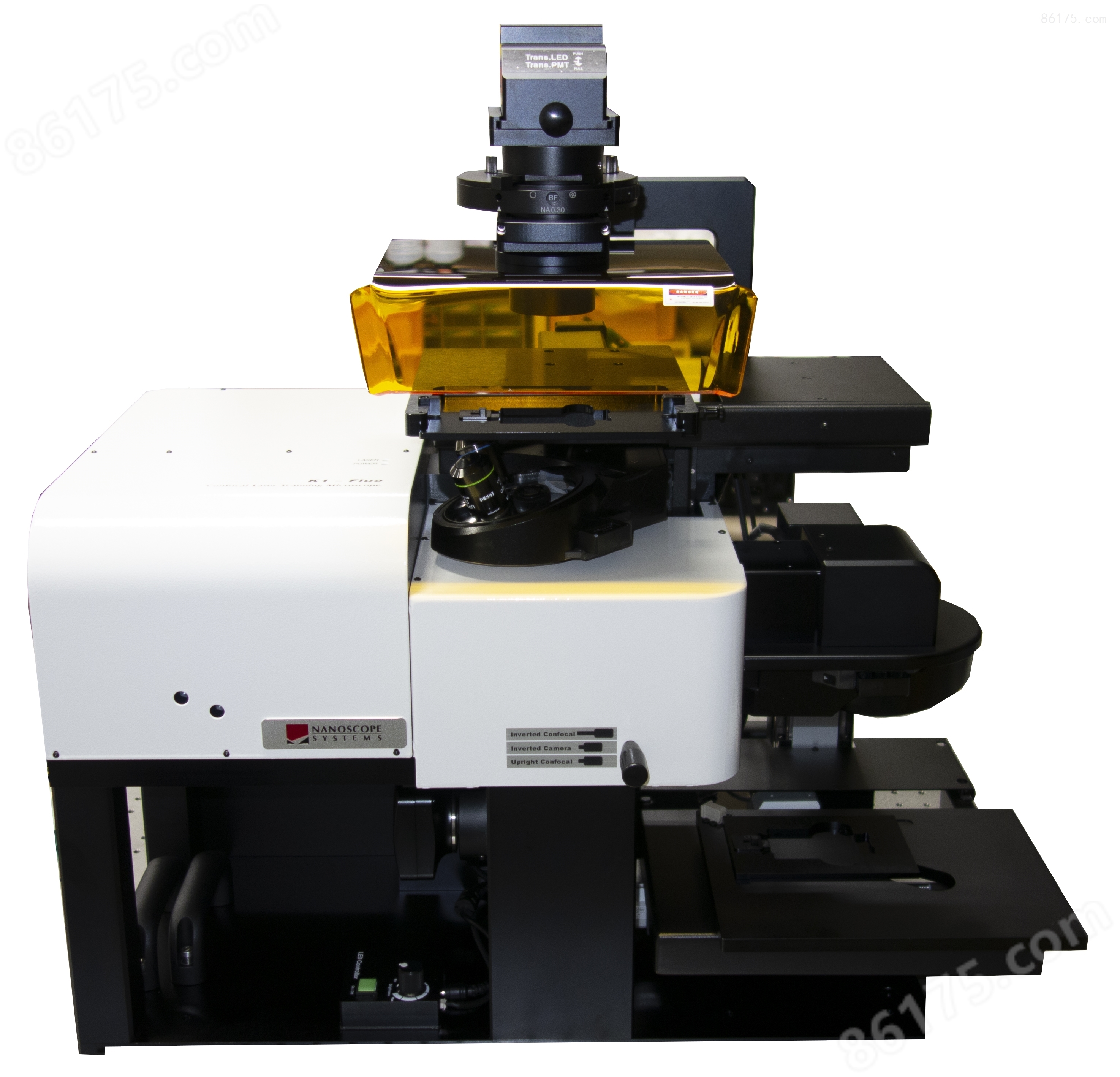
K1 Fluo Laser Fluorescence Confocal Microscope is a multifunctional laser fluorescence confocal microscope developed by Nnaoscope Systems in South Korea for the fields of chemistry, medicine, biology, materials, and more. K1 Fluo can measure 2D and 3D fluorescence images of samples, and also provides Time lapse function. Even if the user is not next to the device, the K1 Fluo system can automatically measure images according to the set program.
Product Details
K1 Fluo Pro Multi functional Research GradeLaser Fluorescence Confocal Microscope
K1 Fluo Pro is a multifunctional laser fluorescence confocal microscope developed by Nanoscope Systems in South Korea for fields such as chemistry, medical, biology, and materials. Excellent optical components, performance, and outstanding cost-effectiveness have attracted a large number of users both domestically and internationally The modular design and open design concept enable the laser control module of K1 Fluo to be compatible with commercial microscope modules from Nikon, Olympus, Zeiss, and Leica
Features & Benefits(Performance and advantages):
1. High definition image quality
2. Compact module size
3. Compatible with various commercial microscope bodies
4. Easy to operate
5. High cost-effectiveness
6. Customizable modules
Main functions:
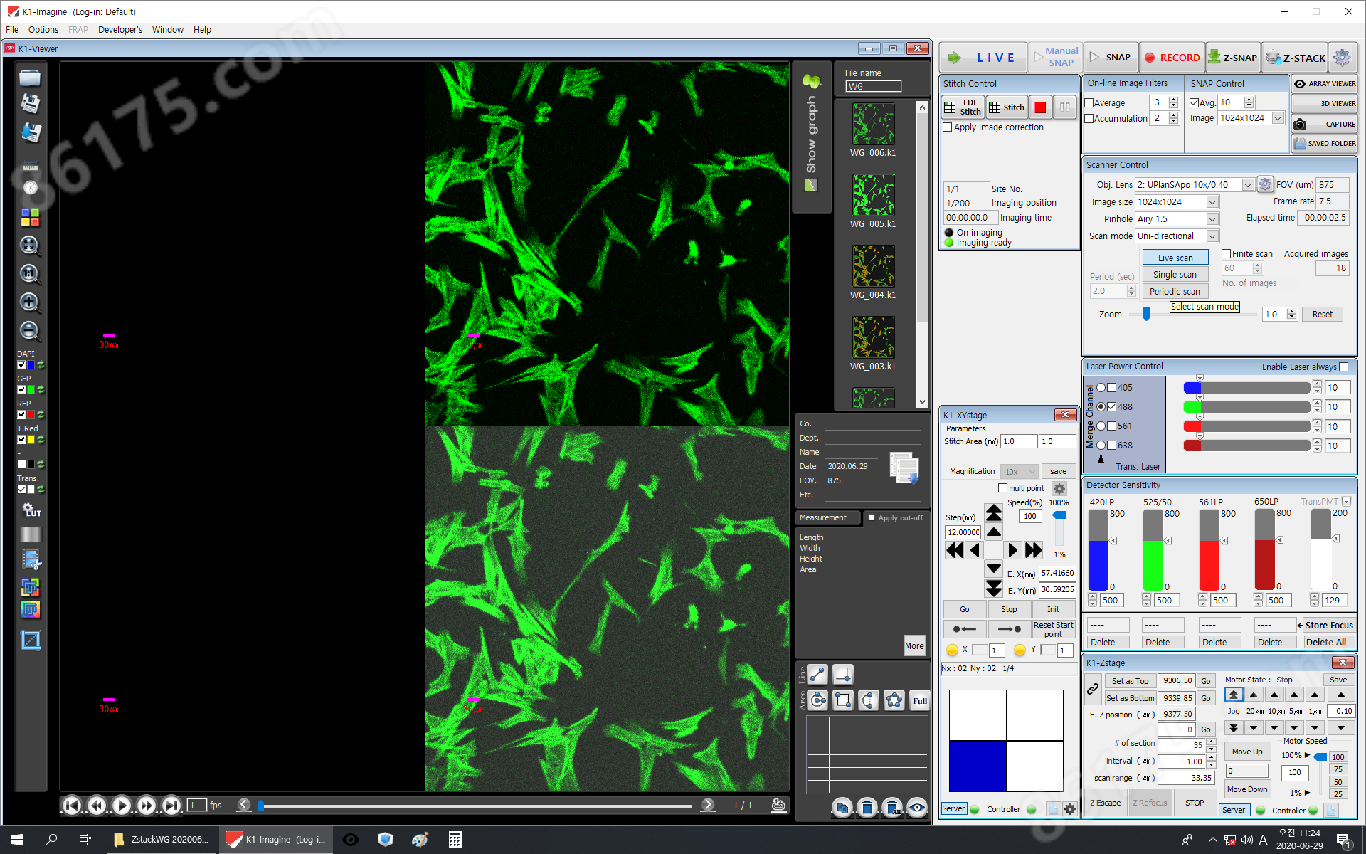
1.Real time mode
In Live mode, high-definition fluorescence images can be obtained by moving the objective lens and adjusting the Detector Sensitivity, and observing changes in the image in real-time.
2. Two dimensional image measurement
When in Live mode, clicking Manual SNAP can measure the two-dimensional image under the current conditions. If you are not satisfied with the current measured image, you can select the laser wavelength for measurement again, adjust the measurement conditions, and measure again to overwrite the previous measurement results .
3. Z-SNAP (2D Depth Image Construction)
By setting the desired scan range, interval, number of section, depth photos of two-dimensional images can be obtained (selecting bright points on each photo and synthesizing them into a two-dimensional photo, which is very suitable when the sample is tilted)
4. Z-STACK (3D Construction)
After setting the desired Scan range, Interval, and # of section, the system reconstructs a 3D image using the measured 2D photos. The K1-3DViewer software allows for observation and analysis of the constructed three-dimensional fluorescence morphology, and enables individual analysis of each measured two-dimensional fluorescence image. The LUT can be adjusted to export intensity values for specific areas, and the three-dimensional dimensions of the fluorescent portion can be measured using the K1 image software.
5. Video function
By measuring multiple consecutive photos and connecting them together, a video can be created. The video size can be adjusted by resetting the frame rate for saving the video.
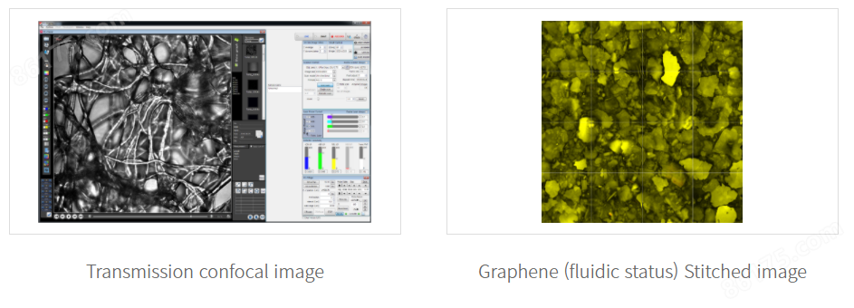
6. Stitch and EDF stitch (flat splicing and depth splicing)
Stitch is a flat stitching function that only scans and stitches images on the same focal plane. EDF stitch is to perform Z-SNAP and Stitch simultaneously at each scanning point (the advantage is that depth images can be seen, but the disadvantage is that it takes a long time)
7. Time lapse
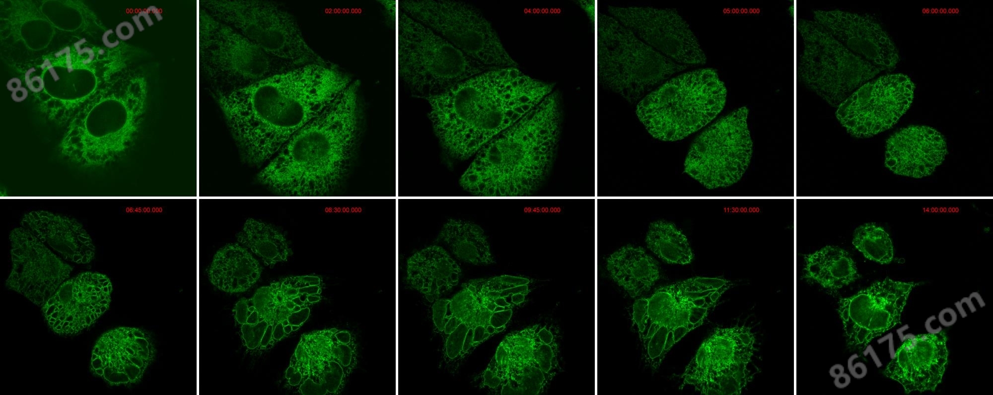
By setting the shooting time interval and the total number of images required, the delay function can be set (especially suitable for tracking cell activity and observing cell division)
8. Automatic Well Plate Scanning
After selecting this function, each corresponding orifice plate coordinate will automatically appear based on the selected orifice plate type. Clicking on each coordinate will automatically move the objective lens to the corresponding orifice plate and perform scanning.
Software(Software):
3-dimensional display (Z-stack)(3D display):
Z-STACK uses the vertical movement of the Z-axis to obtain optical slices and then constructs a 3D image. After selecting the measurement method desired by the user (there are three measurement methods to choose from: Top/bottom, Center/Range, Upper/Lower), the scanning range of the Z-axis (Scan range), the interval of the optical slices (Interval), and the number of optical slices (# of section) are set

Time lapse(Delay function):
Application field:

Specifications(Parameters):
laser module | ||||
laser wavelength |
Basic wavelength |
405, 488, 561nm (7mW each) |
||
Optional wavelength |
445, 473, 514, 532, 637, 640, 660, 685, 705, 730, 785nm (10mW or more), selectable up to four lines |
|||
scanning module | ||||
K1-Fluo HD |
K1-Fluo RT |
|||
scanner |
Two independent flow mirrors |
Resonance scanner and galvanometer |
||
Scanning resolution |
128x128~4095x4095 optional |
128x128~2048x2048 optional |
||
scanning speed |
Variable line frequency of 1~1000Hz |
30fps at 512 x 512 pixels (Bi-scan) |
||
Enlarge range |
0.7x to 7x continuously adjustable |
0.7x~3x continuous |
||
scanning area |
Divide the square of 12.5mm by the magnification of the objective lens (18 fields) |
|||
scanning mode |
xy, xyz, xt, xyt, xyzt |
|||
pinhole |
Automatically switch pinhole size (0.5~10 Airy size) |
|||
weight |
7kg |
|||
Detector module | ||||
detection range |
400-750nm or NIR customizable |
|||
detection range |
standard |
High sensitivity PMT |
||
Low light mode |
Ultra sensitive GaAsP PMT |
|||
Basic Mode |
Six wheel filter turntable with one PMT |
|||
Number of detectors |
Multi ch mode |
Up to 4 PMTs synchronously detected |
||
|
Emission filter |
Automatic filter switching wheel or individual replaceable filter |
|||
Data Depth |
12bit |
|||
weight |
1.5 kg |
|||
Microscope module | ||||
Upright or Inverted |
Digital microscope body (DMB) or commercial microscope body; Nikon, Olympus, Zeiss, Leica commercial microscopes with side holes |
|||
X. Y displacement table |
Electric/manual displacement table; Travel range 115x75 mm (customizable), supports: glass slides, well plates, culture dishes, etc |
|||
Z-drive |
Automatic displacement table |
15mm stroke/250nm small step size |
||
PZT displacement stage (single objective lens) |
400um stroke/1nm step distance |
|||
accessories |
Jog dial DIC (Differential Interference) Sample stage (5 pieces) Magnetic levitation isolation platform optional Cell incubator Objective lens: Supports various specifications of objectives such as air lens, oil lens, water lens, etc Air floating vibration isolation optical platform air compressor |
|||
weight |
12kg |
|||
electronic module | ||||
controller |
electronic device; Semiconductor laser emitter, scanning module, detector module |
|||
Power consumption: 100~240V, 450VA, 50/60Hz | ||||
Weight: 19kg | ||||
PC |
PC |
|||
Windows 10, 64bit | ||||
Power consumption: 100~240V, 900VA, 50/60Hz | ||||


