-
E-mail
szks@xfdoe.com
-
Phone
15205180073
-
Address
Sunshine Building, Shigu Road, Qinhuai District, Nanjing City
Product Categories
- Gel imaging
- Fluorescence quantitative gene amplification instrument
- Imported sample concentration system
- Liquid nitrogen storage device
- Chemiluminescence imaging instrument
- Imported gene amplification instrument
- Flow imaging particle analysis system
- Nano dot delivery system
- High connotation analysis system
Nanjing Xinfeida Optoelectronics Science and Technology Co., Ltd
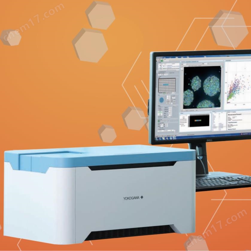
High connotation analysis system
NegotiableUpdate on 01/27
- Model
- Nature of the Manufacturer
- Producers
- Product Category
- Place of Origin
Overview
Through high-throughput 3D imaging for cell detection, the CQ1 high content analysis system provides a new perspective for cell imaging analysis. For many years, the use of confocal microscopy to obtain high-definition 3D imaging has promoted the progress of cell biology research. The high connotation analysis technology combining data analysis and confocal imaging provides a comprehensive solution for cell biology research. CQ1 can achieve high-definition 3D imaging, target recognition, and rapid quantitative analysis of living cells and cell populations. The association between images and data can deepen the interpretation of experimental results and enhance the reliability of data. CQ1 imaging technology reduces phototoxicity to cells and provides temperature controlled imaging chambers ..
Product Details
Cell detection through high-throughput 3D imaging
CQ1New Perspectives on High Content Analysis System Cell Imaging Analysis
Over the years, the use of confocal microscopy to obtain high-definition 3D imaging has driven advances in cell biology research. The high connotation analysis technology combining data analysis and confocal imaging provides a comprehensive solution for cell biology research. CQ1 can achieve high-definition 3D imaging, target recognition, and rapid quantitative analysis of living cells and cell populations. The association between images and data can deepen the interpretation of experimental results and enhance the reliability of data. The imaging technology of CQ1 reduces phototoxicity to cells, and the temperature controlled imaging chamber is like a cell culture box, thus supporting delayed imaging of live cells.
Yokogawa Electric CQ1 is a simple, easy-to-use, and reasonably priced multifunctional confocal microscope. CQ1 has multiple configuration options and supports intelligent integration to achieve fully automated imaging analysis.
High connotation analysis system
Can detect cell spheres, clones, and tissue sections
Unlike flow cytometry, it can detect cells in culture dishes without pre-treatment such as cell detachment
Thanks to the confocal technology of the turntable, 3D images can be quickly and gently obtained
● Supports 4-color excitation light and 10 color emission light; Support bright field/phase contrast imaging
● Support delayed imaging analysis of live cells
Equipped with rich feature extraction capabilities, it facilitates complex cell analysis
● Wide field of view and puzzle function can easily image large samples
Provide analysis functions similar to flow cytometry
Simultaneous image acquisition and analysis (real-time analysis)
● Modular application analysis
The analysis data can be traced back to the original image, and the data and image mutually confirm each other
● Multi in one system, easy to operate
Open platform
Can be used as an image acquisition or analysis device to expand into an integrated detection system
● Can output FCS/CSV/ICE data format readable by third-party data analysis software
● Can be connected to a robotic arm to achieve fully automatic imaging analysis
● Suitable for various cell cultures and sample containers
Small and lightweight desktop devices that do not require a darkroom
Compact and compact, integrating multiple functions into one
■ Confocal scanning unit
Microscope unit
■ Emission filter
Fluorescence imaging illumination (laser)
The multi beam scanning of "wide field confocal micro lens enhanced Nipkow dual disc" can achieve high-throughput 2D/3D imaging, greatly reducing damage to the sample.
High throughput detection of submicron samples is achieved through high-performance objective lenses (super chromatic aberration reduction) and SCMOS cameras with the widest field of view/highest resolution.
Up to 10 emission filters can be installed. Multiple markers can be detected in just one experiment.
Up to 4 solid-state lasers for confocal (fluorescence) imaging can be installed. Multiple fluorescence signals can be detected in just one experiment.


