Which polyacrylamide brands from china are reputable ?
Overview of major polyacrylamide manufacturers in China: 10 brands worth paying attention to09,21,2026 8 views


11,08,20181From:
In a new study, Academician Zihe Rao, researcher Quan Wang, researcher Fei Sun and colleagues from the Institute of Biophysics of the Chinese Academy of Sciences isolated the respiratory super complex of Mycobacterium smegmatis, and used Cryo EM technology to visually observe its three-dimensional structure at a resolution of 3.5 ο. This bacterium has a close genetic relationship with Mycobacterium tuberculosis and is used in many studiesbacteriaThe popular model of species. This detailed structure reveals how electrons are transmitted within cells in a process that has not been observed so far. The relevant research results were published online in the journal Science on October 25, 2018, under the title "An Electron Transfer Path Connections Subunits of a Mycobacterial Respiratory Supercomplex".
Usually, during cellular respiration, the oxidation of energy sources (sugars, fatty acids, and amino acids) is coupled with the reduction of electron acceptors (oxygen, sulfur, nitrate, and sulfate), from which chemical energy can be obtained to synthesize adenosine triphosphate (ATP) and drive cellular reactions. In aerobic cell respiration, this chemical energy is generated by a transmembrane proton gradient called proton motive force (PMF) when the electron donor is transferred through the electron transport chain (ETC) to the terminal electron acceptor, which drives ATP synthesis. In this new study, these researchers revealed a direct correlation between electron transfer between enzymes, representing a new respiratory chain catalytic mode.
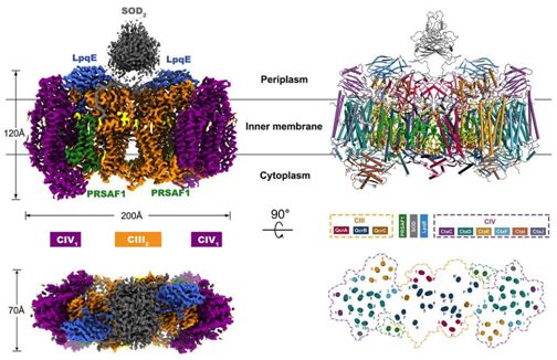
The image is from Science, doi: 10.1126/science. aat8923.
Quinone and cytochrome are two types of electrons in the electron transfer chaincarrierUsed for transferring electrons between larger molecular structures embedded in membranes. Four types of membrane oxidoreductases are involved in electron transfer in the mitochondrial respiratory chain. They include complex I (NADH: ubiquinone oxidoreductase, CI), complex II (succinic acid: ubiquinone oxidoreductase, CII), and complex III (bc1 type panthenol; Cytochrome c oxidoreductase, bc1 type CIII) and complex IV (aa3 type cytochrome c oxidase, aa3 type CIV). Functionally, complex CIII is capable of oxidizing panthenol to ubiquinone and transferring electrons to soluble cytochrome c. The electrons are then transferred to complex CIV, where oxygen is reduced to water. Transmembrane PMF is generated through proton pumping in complexes CI, CIII, and CIV.
In the respiratory chain of prokaryotes, the situation is even more complex. Due to this complexity, the complete electron transfer pathway has not yet been determined in prokaryotic cells. Therefore, it is necessary to understand participationbacteriaThe complete structure of a respiratory chain super complex for electron transfer. In this new study, these researchers extracted and purified this respiratory chain supercomplex from Mycobacterium smegmatis, and visualized its structure using low-temperature electron microscopy technology at a resolution of 3.5 Å. This structure provides important insights into the direct electron transfer mechanism in this respiratory chain supercomplex. The size of this respiratory chain super complex is in the range of 200 × 70 × 120 Å, existing in a symmetrical linear structure, which is different from the previously reported respiratory chain super complexes. In terms of composition, the linear CIV1-CIII2-CIV1 dimer is arranged in such a way that a single complex CIV1 is located on either side of the central complex CIII2 dimer. This information reveals a direct correlation between enzymes during electron transfer, representing a new respiratory chain catalytic mode. This detailed structural discovery has the potential to assist in the discovery of drugs against mycobacteria.
During bacterial cell culture experiments, these researchers used hydrogen peroxide resistant mutant strains of Mycobacterium smegmatis, similar to Mycobacterium tuberculosis. Cultivate these bacterial cells and then isolate their cell membranes according to the previously described method (Microbiology, 2006, 152:823-829, doi: 10.1099/mic.0.28723-0). After bacterial cell culture, collection, and lysis, collect their cell membrane precipitates, and then extract the respiratory chain supercomplex from the cell membrane. They subsequently described the characteristics of this respiratory chain supercomplex using spectroscopic methods, mass spectrometry, and 3,3 '- diaminobenzidine (DAB) staining. In order to identify the hemoglobin group, according to the previous method (Journal of Biological Chemistry, 2015, doi: 10.1074/jbc. M114.624312), they analyzed selected samples by recording spectra before and after the reduction of disulfate. They analyzed the purified respiratory chain supercomplex sample using natural mass spectrometry to study its structure, and used the previously established methodExperimental MethodAnalyze the individual structural components in this respiratory chain super complex.
During the low-temperature electron microscopy analysis, these researchers used uranyl acetate (1%, w/v) to negatively stain 5 μ l of the respiratory chain supercomplex sample of Mycobacterium smegmatis at a concentration of 0.05 mg/ml. Subsequently, images were taken using a FEI Tecnai Spirit microscope running at 120 kV for initial structural model construction. They reconstructed the low resolution structure of this respiratory chain supercomplex by processing 53 microscopic images from negatively stained respiratory chain supercomplex samples. In order to reconstruct the structure of this respiratory chain supercomplex, they manually selected 7600 images from 8200 original images during low-temperature electron microscopy image processing. All images in this study were constructed using PyMOL or UCSF Chimera.
These researchers revealed the low-temperature electron microscopy structure of the respiratory chain supercomplex of Mycobacterium smegmatis CIII-CIV. The range of electron transfer pathways within this respiratory chain supercomplex extends from the oxidation of quinol in complex CIII to the reduction of oxygen in complex CIV. These research results demonstrate a novel forked electron transfer mechanism, ensuring the completion of the quinone cycle (Q cycle, i.e. the net movement of protons through the lipid bilayer) to achieve energy conversion. Oxide dismutase (SOD) directly participates in the assembly of this respiratory chain supercomplex, which can protect it from oxidative damage caused by reactive oxygen species (ROS). The distribution of quinone binding sites also provides a framework for the development of structure based antimicrobial drugs in the future. (Biological Valley)
reference material:
Hongri Gong1,*, Jun Li2,3,*, Ao Xu et al. An electron transfer path connects subunits of a mycobacterial respiratory supercomplex. Science, Published Online: 25 Oct 2018, doi:10.1126/science.aat8923.
Previous111

Which polyacrylamide brands from china are reputable ?
Overview of major polyacrylamide manufacturers in China: 10 brands worth paying attention to
How to Source Reliable Polyacrylamide from Top Manufacturers and Suppliers
Shouxin Advanced Polymer Sciences provides high-performance polyacrylamide (PAM) solutions for water treatment, mining, pulp and paper, and industrial applications.
Polyacrylamide Manufacturer in China | Shouxin Advanced Polymer Sciences Co., Ltd.
Shouxin Advanced Polymer Sciences Co., Ltd. is a leading polyacrylamide (PAM) brand and global supplier based in China.