-
E-mail
info@tvscope.cn
-
Phone
15261811715
-
Address
Super dimensional scenery
Beijing Chaoweijing Biotechnology Co., Ltd
Assisting in the discovery of endothelial cell phagocytosis exacerbates blood-brain barrier damage and its mechanism after ischemic stroke
Date: 2025-10-31Read: 3
Stroke is a major global public health challenge and has become the third leading cause of death and disability in recent years. Although neuronal death is the main pathological mechanism leading to stroke related disability, clinical trials targeting neuronal survival pathways have repeatedly failed, highlighting the urgent need to develop treatment strategies centered on non neuronal pathophysiology. The latest research evidence shows that blood-brain barrier (BBB) dysfunction, as a key pathological mechanism exacerbating ischemic stroke, can lead to neuronal death, edema formation, and poor clinical prognosis. It is not only a key early event in the pathogenesis of ischemic stroke, but also a highly promising therapeutic target.
Neuroscience Bulletin
2025/09
On September 18, 2025, Professor Xu Yun and Professor Wang Chun from Nanjing University jointly published a research paper titled "Endothelial GATAD1 Exacerbates Blood brain Barrier Dysfunction in Ischemic Stroke through Caveolae mediated Transcytosis" in Neuroscience Bulletin, revealing the pathological role of transcription factor GATA zinc finger domain protein 1 (Gatad1) in cerebral vascular endothelial cells (ECs). Gatad1 regulates the CD36-Caveolin-1 (Cav-1) signaling axis, promotes caveolae mediated endocytosis, and ultimately increases BBB permeability and exacerbates brain injury.

Screenshot of the online publication of the paper
In this study, researchers conducted proteomic analysis on mice with middle cerebral artery occlusion (MCAO) model and successfully screened and discovered a significant change in GATA zinc finger domain protein 1 (GATAD1); GATAD1, as an important transcription factor in cardiac development and cardiovascular disease, its role in regulating blood-brain barrier function and ischemic stroke is still unclear. This study found that the protein and mRNA levels of Gatad1 were significantly elevated in the cerebral microvasculature of MCAO model mice; In an in vitro ischemic injury model, it was confirmed that the level of Gatad1 in endothelial cells increased after oxygen glucose deprivation (OGD).

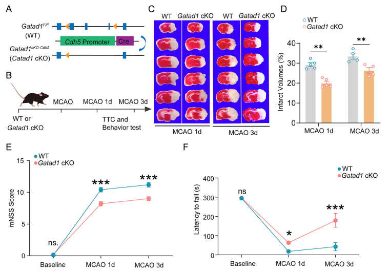
In order to explore the pathological role of Gatad1 in vascular endothelial cells after ischemic stroke, researchers constructed transgenic mice with specific knockout of Gatad1. Further research found that after MCAO in Gatad1 knockout mice, TTC staining showed a 32% reduction in cerebral infarction volume compared to wild-type mice (P<0.01), and behavioral studies showed improvement in neurological deficits. This suggests that the reduction of Gatad1 has a certain neuroprotective effect.
As a key component of the blood-brain barrier, endothelial cells are interconnected through tight junctions (TJs) to limit molecular diffusion. The transmission electron microscopy results showed that there were gaps in the tight connections of the ipsilateral blood vessels in the penumbra area of ischemic stroke, and adjacent endothelial cell membranes lost contact, which were not observed in healthy or contralateral cortex; The proportion of abnormally tight junctions in Gatad1 knockout mice was significantly lower than that in wild-type mice on day 1 after MCAO, but there was no significant difference between the two at 6 hours after MCAO. To further validate the protective effect of Gatad1 knockout on MCAO, the researchers employedMicro scale two-photon microscopy imaging technology for super dimensional organismsThe dynamic changes of the semi dark band blood-brain barrier (BBB) were recorded before and during stroke progression. The results showed that tracer leakage in WT mice gradually increased within 24 hours, while in Gatad1 knockout mice, it reached its peak 12 hours after MCAO and began to decrease 12-24 hours later.
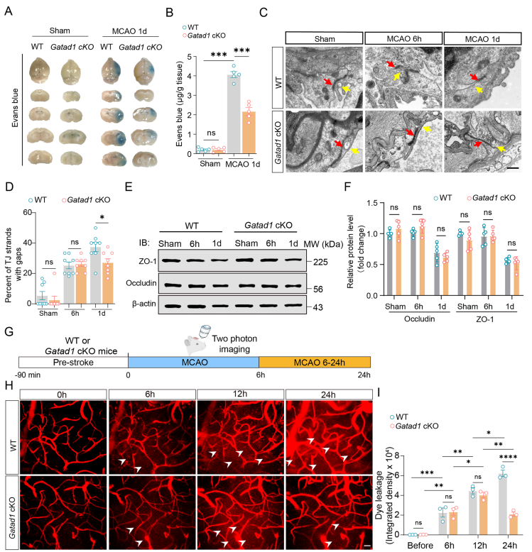
It is worth noting that previous studies have shown that the cross cellular mechanism of blood-brain barrier function is impaired as early as 6 hours after stroke, and structural defects appear in tight junctions at least 24 hours after MCAO. This indicates that blood-brain barrier disruption within 1 day after MCAO is mainly caused by cross cellular damage to the blood-brain barrier. Therefore, the results of this study indicate that Gatad1 deficiency specific to vascular endothelial cells can rescue the disruption of the blood-brain barrier in mice after MCAO through a cross cellular mechanism.
[Reference]
Fan L, Liu H, Li S, et al. Endothelial GATAD1 Exacerbates Blood-brain Barrier Dysfunction in Ischemic Stroke through Caveolae-mediated Transcytosis. Neurosci Bull. Published online September 18, 2025. doi:10.1007/s12264-025-01507-z