- Phone
-
Address
Building 8, the Pearl River Moore International Building, Changping District, Beijing
Product Categories
Beijing Ruike Zhongyi Technology Co., Ltd
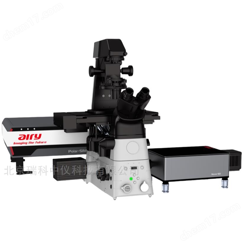
Ultra high resolution and rotary confocal microscopy imaging system
NegotiableUpdate on 07/07
- Model
- Nature of the Manufacturer
- Producers
- Product Category
- Place of Origin
Overview
Ultra high resolution and rotating disc confocal microscopy imaging system $n structured light SIM ultra high resolution and rotating disc confocal microscopy imaging system applied in biomedical field
Product Details
Ultra high resolution and rotary confocal microscopy imaging system
Ultra high resolution and rotary confocal microscopy imaging systemIntegrating ultra-high resolution imaging and fast 3D dynamic observation, utilizing cutting-edge optical design and intelligent image processing technology, we provide microscopy imaging solutions for fields such as life sciences and materials science. The system is equipped with a new solid-state laser group, covering405nm-640nmMulti wavelength excitation combined with high numerical aperture objective lens(100X/1.49 NAOil mirror), achieve200nmUltra high resolution imaging can clearly capture the fine structure of organelles and the distribution of nanoscale biomolecules.

The confocal module of the turntable adopts a high-speed dual turntable design and is equipped with5000rpmAsynchronous rotating disk and micro lens array for increased luminous flux3At the same time, the suppression rate of stray light outside the focal plane is increased to95%Above, achieve every second30Three dimensional imaging of frames, adapted for observing dynamic processes of living cells, such as cell migration, vesicle transport, and other rapid physiological activities. The system has a built-in dual-mode imaging switching function, allowing users to switch between ultra-high resolution static observation and turntable confocal dynamic tracking modes with just one click according to their needs, balancing spatial and temporal resolution.
In terms of hardware configuration, adoptsCMOSCamera(6.5μmPixel size,95%Quantum efficiency) andpiezoNano displacement platform (positioning accuracy)±1nm)Combined with an adaptive optical correction system, it effectively compensates for aberrations caused by sample thickness, ensuring the quality of deep imaging of thick samples. In terms of software, integrationAIAuxiliary image analysis module, supporting automatic cell counting3DStructural reconstruction, fluorescence co localization analysis, and other functions are providedPythonThe interface facilitates the development of personalized data analysis processes.
The system has multiple sample compatibility and can be equipped with live cell culture devices (temperature control accuracy)±0.5℃,CO₂Concentration control±0.1%)And optical tweezers module, meeting the diverse research needs from fixed sections to live cells, from single-molecule localization to tissue 3D imaging. The overall design follows the principles of ergonomics, with a modular architecture that supports functional expansion. It is equipped with an anti vibration optical platform and a low-noise shielding design, providing a stable experimental environment for long-term high-sensitivity imaging. It is an ideal tool for conducting cutting-edge biomedical research.
Similar Product Recommend