- Phone
-
Address
3rd Floor, Building B1, Science and Technology Oasis, No. 579 Xingmei Road, Minhang District, Shanghai
Product Categories
Shanghai Yuyan Scientific Instrument Co., Ltd

Ultra high frequency small animal ultrasound imaging system
NegotiableUpdate on 05/07
- Model
- Nature of the Manufacturer
- Producers
- Product Category
- Place of Origin
Overview
The ultra-high frequency small animal ultrasound imaging system is equipped with advanced high-resolution imaging platforms and high-frequency probes, and integrates cutting-edge ultrasound imaging technology, resulting in high image quality.
Product Details
Ultra high frequency small animal ultrasound imaging system
Ultra high frequency small animal ultrasound imaging systemEquipped with a high-resolution imaging platform and high-frequency probe, and integrated with ultrasound imaging technology, the image quality meets the standard
Product Details
Sonorover UR820Ultra high frequency small animal ultrasound imaging systemEquipped with advanced high-resolution imaging platform and high-frequency probe, and integrated with ultrasound imaging technology for image quality. Fully meet the needs of biomedical research fields such as cardiovascular disease, abdominal organs, tumors, and embryos in small animals, and assist researchers in continuously exploring unknown paths.
Product Features
256 channels: The system uses 256 channels for better imaging performance and superior image quality
120MSPS: The sampling rate of the ultrasonic analog signal ADC chip can reach 120MSPS
256 elements: High density 256 element ultrasound imaging probe, meeting the needs of high-performance ultrasound imaging
50 μ m: The image resolution can reach 50 μ m, providing richer and more accurate visual information

Advanced ultrasound probe

High density 256 element ultrasonic imaging probe, high-performance piezoelectric material combined with multi-layer matching technology, low capacitance ultrasonic dedicated cable, supporting multiple frequency probes
| model | frequency range | application |
|---|---|---|
| L50-18A | 18-50MHz | Mice, zebrafish/blood vessels, embryos, skin |
| L32-14A | 14-32MHz | Mouse, rat/abdomen, reproductive, cardiovascular, embryonic, tumor |
| L21-9A | 9-21MHz | Rats, rabbits/blood vessels, embryos, skin |
| L14-6A | 6-14MHz | Cat, dog/cardiovascular, abdominal, embryo |
| L12-4A | 4-12MHz | Pigs, cows/cardiovascular, abdominal, embryonic |
Image enhancement function
One click optimization of contrast and resolution, improving diagnostic and scientific accuracy, enhancing image clarity and detail representation
Myocardial strain imaging
An important tool for cardiac research: myocardial strain imaging can non invasively and real-time evaluate myocardial function, helping researchers to explore the pathogenesis, drug efficacy, and gene function of cardiovascular diseases in depth
Comprehensive assessment of myocardial function: Strain imaging provides multidimensional strain analysis, helping to comprehensively understand the contraction and deformation of the myocardium in different directions, and improving the accuracy of diagnosis
Quantitative analysis: Strain imaging can quantify myocardial deformation and provide accurate numerical parameters for cardiac function
Detecting synchronicity: By measuring the deformation of different myocardial segments, strain imaging can reveal the asynchrony of myocardial contraction

High frame rate contrast imaging
Higher temporal resolution: By increasing the frame rate of imaging, higher temporal resolution can be obtained, which can display more microvascular architecture and vascular perfusion details more intuitively and clearly
Fast dynamic capture: Record the dynamic changes of contrast agents in the bloodstream at ultra-high frame rates. The maximum frame rate can reach 120 frames per second
Microcirculation imaging: providing precise microvascular structure and blood flow perfusion information
Accurate lesion assessment: achieving early detection and quantitative analysis of small lesions

3D/4D imaging
Cardiovascular research: studying the structure, movement, and blood flow changes of small animal hearts, suitable for model studies of congenital heart disease, cardiomyopathy, and other conditions
Tumor Research: Observing Tumor Blood Supply, Angiogenesis, and Treatment Response
Detecting synchronicity: monitoring heart and organ function during the development of small animal embryos
Drug development: Evaluating the impact of drugs on cardiovascular or organ function

Super resolution ultrasound imaging
Tumor microvascular imaging: Abnormal microvascular growth is one of the characteristics of cancer, and super-resolution ultrasound imaging can be used for basic research and clinical management of cancer, such as early detection, diagnosis, and treatment response evaluation. Early diagnosis can be achieved through changes in microvascular morphology when the tumor volume is still small
Brain microvascular imaging: provides imaging coverage and micrometer level spatial resolution of the brain, while measuring the blood flow velocity of individual blood vessels. For example, in stroke research, early detection of microvascular blood flow changes in the ischemic penumbra can guide thrombolytic therapy. Observing vascular damage in amyloid plaque areas in Alzheimer's disease models and studying disease mechanisms

High frequency photoacoustic imaging
High frequency ultrasound clearly displays tissue structure through fine spatial resolution, while photoacoustic technology captures molecular functional information such as blood oxygen levels based on photoacoustic effects. The advantages of the two complement each other and can achieve high-resolution, multidimensional imaging of tissues
High resolution: Combining optical and ultrasound technology to provide high-resolution images of tissue structure and function
Deep penetration: Compared to pure optical technology, photoacoustic imaging can penetrate deeper tissues and is suitable for a wider range of examinations or studies
Functional imaging: achieving functional imaging of oxygen saturation, vascular function, and molecular level, supporting precision medicine research
Multi mode fusion: can be combined with ultrasound imaging technology to obtain more tissue information and improve diagnostic accuracy
| parameter indicators | numerical value |
|---|---|
| Host collects real-time frame rate | ≥ 10000 frames per second |
| pulse frequency | ≥50Hz |
| Adjustable wavelength range | Two frequency bands, 680-970nm and 1200-2000nm |
| Minimum precision of laser adjustment | 1nm |
| Photoacoustic acquisition frequency | ≥ 50 frames per second |
| Multispectral imaging | Excitation of ≥ 5 arbitrary wavelengths and composite photoacoustic imaging |

Functional Brain Imaging (fUS)
FUS can detect brain functional activity through neurovascular coupling mechanisms, and can be applied to various animals such as mice, rats, and non-human primates to assist in the study of cerebrovascular related diseases and mechanisms. It can also be applied to various animal models related to neurological or psychiatric disorders, neuropharmacological research, new drug screening to find new disease-related biomarkers, and so on. The application in preclinical research is very extensive.

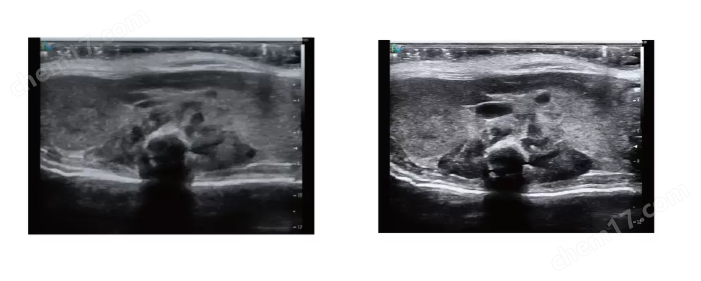
Imaging effect display


