- Phone
-
Address
906, Building A, Shangao Century Center, Xisanqi, Haidian District
Shilian Boyan (Beijing) Technology Co., Ltd

Living Cell Imaging Fluid System
NegotiableUpdate on 05/06
- Model
- Nature of the Manufacturer
- Producers
- Product Category
- Place of Origin
Overview
Live Cell Imaging Fluid System $r $n is a fully automated culture medium and reagent transfer system used for live cell imaging. It conforms to the standard ANSI/SBS packaging and is suitable for the top and bottom of the stage; #174; incubator. You can easily set up various experimental plans on your PC, and the system will automatically execute the pipetting process you are currently manually performing. The pump head and other liquid receiving components are disposable.
Product Details
Living Cell Imaging Fluid System
feature
1. Easy to program, from quick medium replacement to gentle infusion. Researchers only need to press“start”You can start fully automatic pipetting andFocus on cell observation.
2. Can slowly add reagents(uL/minThis is difficult to achieve with traditional manual pipetting. Controlled power supply speed can generate stable images.
3. The gradient mode can be gradually changed2Control the concentration by adjusting the flow rate of the infusion pump. (Only applicable to)CEIM-0200Series)
4. many 4 The types of reagent addition pumps have different flow rates. Please contact us for more information.
*When using a peristaltic pump, there may be a certain degree of pulsation.
specification
model |
CEIM-010X |
CEIM-020X |
Media feed/Waste pump flow rate |
3 ~ 10milliliter/minute |
3 ~ 10milliliter/minute |
Reagent addition pump flow rate |
0.45 ~ 7milliliter/minute |
0.1ul ~ 7milliliter/minute |
Gradient mode |
× |
○ |
size |
100 × 240 × 100 mm |
100 × 240 × 100 mm |
Supported operating systems |
Windows 7 And higher versions |
Windows 7 And higher versions |
Appropriate diameter of culture dish |
35millimeter |
35millimeter |
Application example
Due to the fact that the sample addition does not interfere with imaging, even with high magnification/High numerical aperture microscope(60X,NA1.35)You can also clearly observe the images before and after adding the reagent sample.
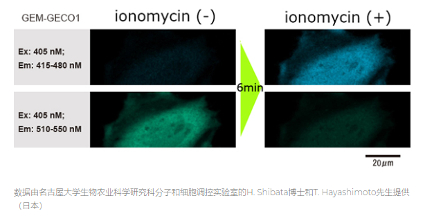
1. Translate into EnglishHeLaCells were fed into glass bottomed culture dishes and transfected with genes.
2. After incubating for one day, add during photography1mlReagents stimulate genes.
3. Observed within the cellGEM-GECO1The change in fluorescence wavelength (whenGEM-GECO1When chemically bonded with calcium ions, the color of the fluorescence wavelength changes from green to blue.
[Experimental conditions]
Microscope: Inverted confocal laser microscope objective lens
Magnification factor:60times
Objective numerical aperture:1.35
Cell: HeLa cell
Reagent: Leucomycin
system configuration
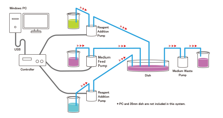
Living Cell Imaging Fluid System


