-
E-mail
rliu_1@sypi.com.cn
- Phone
-
Address
Building 1, Huadian Intelligent Network Industrial Park, No. 28 Siwei Road, Dongli Development Zone, Tianjin
Solution
Tianjin Sanying Precision Instrument Co., Ltd
The ancients called pears the "king of all fruits". In the golden autumn of September, when the weather is dry and everything is dry, pears are a beloved fruit among people. However, pears with many benefits can also get sick, and cork disease is one of the most harmful physiological diseases in pear fruit, causing significant economic losses to fruit farmers every year. To this end, Cui Zhenhua, Wang Nannan, and others from Qingdao Agricultural University, based on previous research, used three precision X-ray micro CT (nanoVoxel-3000) to select pear fruits of the "Akizuki" variety, which is prone to cork disease, for scanning analysis, observed the microstructure of each part, further clarified the changes in fruit texture between healthy and cork disease fruits, and conducted further research on the mechanism of cork disease occurrence.
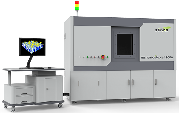
Sanying Precision NanoVoxel-3000 Micro CT
Pear fruit appears:
Scanning analysis was conducted on healthy pear fruits and pear fruits with cork disease, and it was found that the surface of healthy pear fruits was smooth, while the surface of pear fruits with cork disease had obvious collapse pits. However, these pits were not obvious to the naked eye.
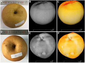
(A)Healthy pear fruits (B, C) are rendered in gray and yellow after X-ray scanning
(D) Pear fruit with cork disease (E, F) The results of rendering the pear fruit with cork disease in gray and yellow after X-ray scanning
Distribution analysis of solid wood bolt spots on pear fruit:
Healthy pear fruit andCork diseasePear fruits were stained with safranin solution, and it was found that cellulose (red dots stained with safranin) was presentCork diseaseThe content in pear fruit is higher than that in healthy pear fruit. Based on X-ray CT scanning analysis, observe the transverse and longitudinal sections of two types of pear fruits,Cork spot(i.e. larger and stronger pores) mainly distributed in the flesh near the fruit peel andCork diseaseThe end of the calyx of pear fruit. Further confirmed the effectiveness of X-ray CT scanningCork diseaseReliability of microstructure analysis of pear fruit.
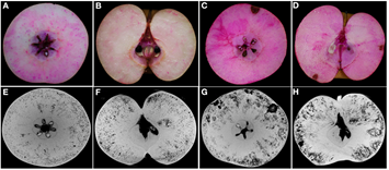
(A,B)The horizontal and vertical sections (C, D) of healthy pear fruit stained with safranin solution are the horizontal and vertical sections (E, F) of pear fruit stained with safranin solution, respectively. The horizontal and vertical sections (G, H) of healthy pear fruit extracted from X-ray scanning data are the horizontal and vertical sections of pear fruit stained with safranin solution, respectively
Analysis of Pore Structure in Pear Fruit:
Using X-ray micro CT technology, compare the threshold data of healthy pear fruits and pear fruits with cork disease after scanning, and analyze the pore structure. The results showed that the porosity of pear fruit with cork disease (9.37%) was significantly different from that of healthy pear fruit (3.52%).
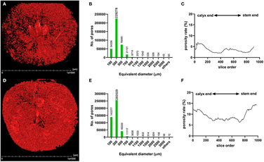
(A,D)The pore structures (B, E) of healthy pear fruit and cork diseased pear fruit are respectively represented by histograms of the number and size of pores in healthy pear fruit and cork diseased pear fruit (C, F). The porosity analysis of healthy pear fruit and cork diseased pear fruit is shown from the end of the fruit calyx to the end of the stem
Analysis of Pore Connectivity in Pear Fruits:
Perform pore connectivity analysis on HRA (high risk area of cork disease) and MRA (medium risk area of cork disease) of healthy pear fruits and cork disease pear fruits. The reconstructed three-dimensional (3D) network model shows that in the pear fruit with cork disease, the pore channels are highly branched and connected; In healthy fruits, there are fewer branches and lower connectivity of pores.
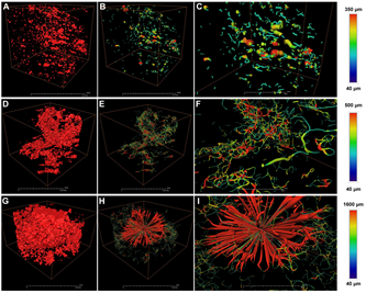
(A)Pore analysis of HRA (high risk area of cork disease) and MRA (medium risk area of cork disease) in healthy pear fruits(B) The reconstructed 3D network model of Figure A (C) and the close-up of Figure B (D, G) show the pore analysis of MRA and HRA of pear fruit with cork disease (E, H), respectively. The reconstructed 3D network models of Figure D and Figure G (F, I) are the close-up of Figure E and Figure H, respectively
Analysis of Pear Kernel Structure and Pore Connectivity:
XThe analysis of the nucleus structure reconstructed by X-ray micro CT scanning shows that the surface of healthy pear nucleus is smooth, while the nucleus of pear with cork disease is damaged and rough, and nucleus deformation only occurs in pear fruit with cork disease. Compared with the long and narrow pores of healthy pear kernels, the pores of pear kernels with cork disease have a highly branched network structure.
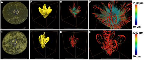
(A,E)The sliced images of healthy pear fruit and pear fruit with cork disease after X-ray scanning (B, F) are the reconstructed 3D models of healthy pear fruit nucleus and pear fruit nucleus with cork disease (C, G), and the 3D network models of Figure B and Figure F (D, H) are the close-up images of Figure C and Figure G, respectively
Microstructure analysis of pear flesh:
The flesh tissues of healthy pear fruit and pear fruit with cork disease were taken separately for MRA (medium risk area of cork disease). The high-resolution (0.5 μ m) scan results of X-ray micro CT showed that the pore size of cork disease pear flesh (87 µ m) was four times that of healthy pear flesh (22 µ m), and the difference in pore size between the two was significant.
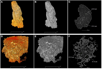
(A,B)Reconstructed 3D images of yellow and gray flesh from MRA (medium risk area of cork disease) of healthy pear fruit (C) X-ray scan segments of healthy flesh (D, E) Reconstructed 3D images of yellow and gray flesh from MRA of cork disease pear fruit (F) X-ray scan segments of cork disease flesh
Conclusion:
High resolution micro CT scanning is a powerful tool for analyzing the microstructure of pear fruit with cork disease. Unlike previous studies, researchers have improved their understanding of the microstructure characteristics of pear fruit, nucleus, and flesh from a new perspective. Compared to healthier pear fruits, pear fruits with cork disease have higher porosity and connectivity of branching pores, which in turn blocks the transport of certain nutrients. This may be one of the reasons why pear fruits suffer from cork disease.
All along, Sanying Precision has been dedicated toXrayCTImaging technology and product development are committed to providing users with new methods and tools to solve problems, and will continue to provide stronger assistance to researchers in the future!