-
E-mail
ustc_sc@163.com
-
Phone
18020740586
-
Address
Room 1503, Building A04, Software Park Phase III, Jimei District, Xiamen City, Fujian Province
About Us
Opu Tiancheng (Xiamen) Optoelectronics Co., Ltd
Date: 2024-01-03Read: 2
Introduction
Winter is a high-risk period for respiratory infections, and currently, respiratory diseases are prevalent in ChinaCaused by multiple pathogenscause, including bacteria and mycoplasma、influenza viruswithAnd coronavirus, a new type of virus, which caused a global pandemic. Considering the respiratory tractdiseaseHighly contagious and contagiousfast speedThe characteristics,And differentThere are significant differences in symptoms and treatment of respiratory infections caused by pathogens, so early detection and diagnosis are effective in controlling respiratory tract infectionsDisease SpreadImportant means. Although traditional separation and cultivation methods have significant advantages in accuracy and authority, they are no longer able to meet the practical needs of current clinical testing due to their cumbersome operation, long time consumption, and involvement of biosafety risks. At present, the main detection methods used include nucleic acid testing and immunological testing, but these methods require professional laboratory equipment, operators need to receive professional training, the cost is relatively high, and the detection cycle is also relatively long,Unable to meet the rapidly increasing respiratory tractDisease samplesThe urgent need for large-scale on-site testing,Therefore, there is an urgent need to develop new respiratory virus detection technologies.
When light scatters on a substance, the scattered light with a change in wavelength is called Raman scattering. The reason for the change in wavelength is that after energy transfer occurs between photons and the irradiated substance molecules, the vibrational energy levels of the molecules change,When photons undergo Raman scattering on different substances, the energy transferred varies, allowing for specific identification of the type of irradiated substance. And surface enhanced Raman spectroscopy(SERS)Technology is based on ordinary Raman scattering to irradiate the material being irradiatedAdsorption or connectionOn the rough surface of precious metals, the Raman signal intensity is greatly enhanced. On the basis of the inherent advantages of non-destructive analysis, unaffected by water composition, no need for complex sample processing, fast detection speed, and on-site analysis using portable instruments in ordinary Raman scattering, SERS technology has higher sensitivity and can achieve single-molecule detection level under certain conditions. By analyzing the biological samples of patients, this technology can quickly detect specific molecular markers, thereby determining the causes of respiratory diseasesSpecially causedpathogeny. Helps medical professionals make accurate diagnoses earlier and provide patients with more timely and personalized treatment plans. By shortening the diagnosis time and reducing the tedious process of traditional examinations, rapid testing methods are expected to play a key role in preventing and controlling infectious diseases, improving medical efficiency, and reducing medical burden, providing more effective support for the treatment of respiratory diseases.
Application of SERS technology in detection of COVID&-19
Coronavirus is the seventh coronavirus capable of infecting human beingsandHaving&Due to its extremely high infectivity, it is particularly urgent to develop a new detection method that is ultra fast and highly sensitive. LEONG et al. designed a handheld new type coronavirus detector based on SERS technology. The instrument integrates three types of chips internally, each carrying three sets of SERS probe molecules: 2-mercaptobenzoic acid, 4-mercaptopropydine, and adenosine triphosphate. These SERS probe molecules are immobilized on the surface of silver nanoparticles. During the detection process, the subject breathes into the device for about 10 seconds. Because the new coronavirus biomarker in the breath reacts with the sensor chemically, the compound after reaction can be characterized by the change of SERS signal. For on-site detection of new coronavirus in 501 people in hospitals and airports, LEONG et al. showed that the false negative rate of this detection method was 3.8%, and the false positive rate was 0.1%. This has comparable accuracy compared to polymerase chain reaction detection, and is lower in cost, with a detection time of only 5 minutes. It is worth noting that this instrument can not only detect changes in the types of volatile organic compounds from biological sources, but also be used to screen for other types of respiratory virus infections.
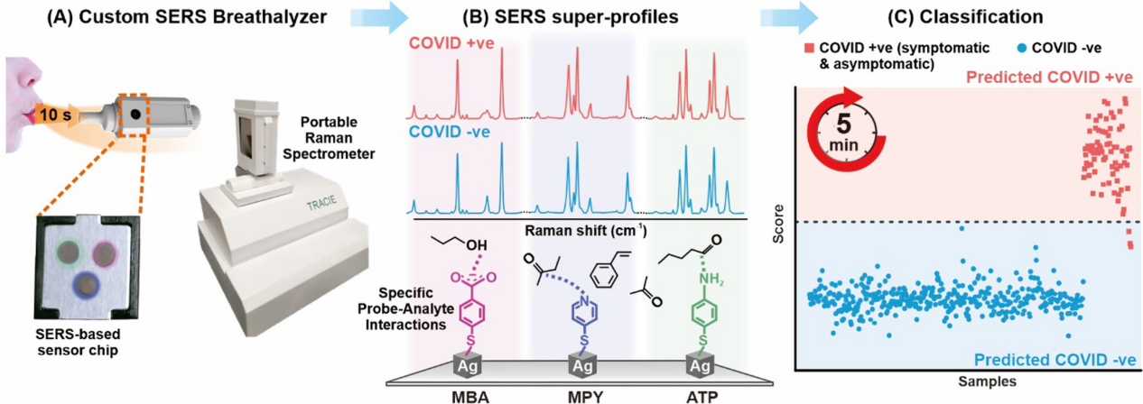
Figure 1 Scheme design of identifying new coronavirus disease virus positive individuals based on respiratory volatile organic compounds.
Application of SERS technology in influenza virus detection
Influenza virus has high infectivity and mutation rate, which may cause seasonal epidemics and even global pandemics, posing a serious burden on public health. Therefore, there is an urgent need to develop an accurate and rapid method for detecting influenza virus, in order to contain its spread through timely and accurate testing before the outbreak of influenza. Chen et al. developed an adaptive sensor based on dual-mode surface enhanced Raman scattering (SERS), which can accurately diagnose and distinguish new&crown&disease&virus&A H1N1 influenza at the same time. In this technology, DNA aptamers that selectively bind to SARS-CoV-2 and H1N1 influenza A are co immobilized on a popcorn shaped gold nanosubstrate. Raman reporter genes (Cy3 and RRX), connected to the ends of DNA aptamers, can generate strong SERS signals in the nanogaps of gold nanosubstrates. At the same time, the internal standard Raman reporter gene (4-MBA) was immobilized on a gold nano substrate along with the aptamer DNA to reduce measurement errors caused by environmental changes. When SARS-CoV-2 or influenza A virus approaches the gold nanosubstrate, the corresponding DNA aptamers selectively separate from the substrate due to the significant binding affinity between the DNA aptamers and the virus. Therefore, as the concentration of the target virus increases, the corresponding SERS intensity gradually decreases. This SERS based DNA aptamer sensor can quickly determine whether a suspected patient is infected with SARS-CoV-2 or influenza A, while quantitatively evaluating the concentration of the target virus with high sensitivity, unaffected by cross reactivity.
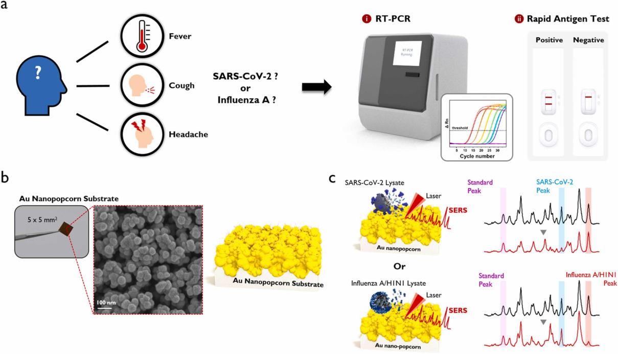
Figure 2(a) Symptoms of SARS-CoV-2 or influenza A infection, as well as RT-PCR and rapid antigen kits used for diagnosis;(b) PopcornZhijinPhotos and SEM images of nano substrates;(c) Dual aptamer fixed popcorn for virus detectionformGold NanobaseThe working principle.
Application of SERS technology in bacterial infection detection
bacterial infectionyesThe main cause of death claims 6.7 million lives annually in both developed and developing countries. The treatment costs for these infections are also high, accounting for 8.7% of annual healthcare expenditures, or $33 billion, in the United States alone. The current diagnostic methods require sample culture to detect and identify bacteria and their antibiotic sensitivity, which is a slow process that may take several days even in * *'s laboratory. Broad spectrum antibiotics are usually prescribed while waiting for the culture results, and according to data from the Centers for Disease Control and Prevention, over 30% of patients receive unnecessary treatment. Therefore, there is a need for new methods for rapid, non cultured diagnosis of bacterial infections, in order to prescribe targeted antibiotics earlier and help alleviate antibiotic resistance. Raman spectroscopy has the potential to identify bacterial species and antibiotic resistance, and when combined with confocal spectroscopy, it can mechanically analyze individual bacterial cells. Different bacterial phenotypes have unique molecular compositions, resulting in subtle differences in their corresponding Raman spectra. However, due to the low Raman scattering efficiency (~10)?8Scattering probability), these subtle spectral differences are easily masked by background noise. Therefore, high signal-to-noise ratio is required to achieve high recognition accuracy, usually requiring a long measurement time, which hinders the development of high-throughput single-cell technology. In addition, a comprehensive dataset is required for a large number of clinically relevant species, strains, and antibiotic resistance patterns, which have not been collected in studies focused on distinguishing species isolates or antibiotic sensitivity. Ho et al. addressed this challenge by combining SERS technology with convolutional neural network training, classifying bacterial SERS spectra through separation, empirical treatment, and antibiotic resistance.
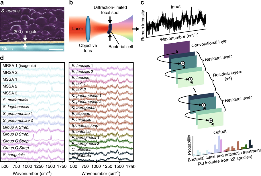
Figure 3Convolutional neural networks are used forSERSIdentify bacteria in the spectrum.
Summary
With the advancement of SERS technology, there are currently three main application ideas in the detection field: (1) attaching specific nucleic acid sequences and antibodies to the surface of nanoparticles to construct SERS tags, thereby achieving accurate detection of respiratory viruses in complex body fluids; (2) By utilizing novel enhanced substrate preparation techniques, the size of the "hotspots" between nanoparticles is more suitable for virus particles, improving detection efficiency and reducing costs, while effectively avoiding interference from biological backgrounds such as saliva and blood on detection results; (3) Combined with other detection techniques, such as SERS chromatography, SERS enhanced substrates are assembled onto optical fibers as highly sensitive detection sensors. In addition, the combination of SERS and plasmonic sensing can be used for high-sensitivity quantitative detection of biomolecule interactions. SERS technology overcomes the weakness of ordinary Raman spectroscopy signals and provides difficult to obtain structural information.
SERS technology has made significant progress in the detection of respiratory disease pathogens. However, in order to comprehensively and deeply study and diagnose respiratory viruses, researchers usually choose to use confocal microscopy Raman spectroscopy. This instrument combines the characteristics of laser confocal microscopy and Raman spectroscopy, providing unique advantages for SERS detection of respiratory disease pathogens:
High spatial resolution: Confocal Raman spectrometer has high-resolution imaging capability, which can observe the structure and organization of samples at the microscopic scale. In respiratory disease pathogen detection, this high spatial resolution can help researchers accurately locate and observe tiny virus particles or cellular structures.
Depth information acquisition: Confocal Raman spectroscopy can achieve three-dimensional imaging, providing researchers with depth information inside the sample. This is crucial for understanding the distribution and interactions of pathogens at different cellular levels, and helps to gain a more comprehensive understanding of the development process of respiratory diseases.
Real time monitoring: The confocal microscopy Raman spectrometer has real-time monitoring function, which can track the dynamic changes of pathogens in the sample. In the study of respiratory diseases, real-time monitoring helps researchers understand the lifecycle of viruses, infection processes, and the effectiveness of treatment interventions.
Reduce background interference: Confocal Raman spectrometer can reduce background signal interference by precisely controlling the focus. This is particularly important for conducting high-sensitivity SERS detection in complex biological samples to ensure the accuracy and reliability of the detection results.
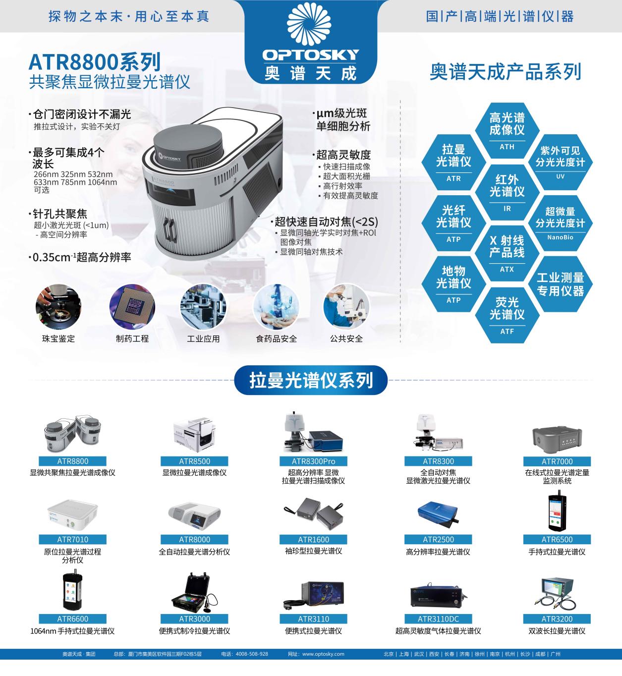
AOPU Tiancheng ATR8800 series micro Raman spectrometerBy integrating up to four lasers and combining the advantages of both microscopy and Raman spectroscopy, the micro Raman detection platform enables "what you see is what you measure". The visualized and precise positioning Raman detection platform allows observers to detect Raman signals of different surface states on the sample and synchronously display the micro area morphology of the detected location on the computer, greatly facilitating Raman micro area detection.
ATR8800The entire series can perform fully automatic focusing and scanning, with one click operation, batch experiments, uniformity scanning, etc., without waiting, and can obtain highly reliable scanning imaging Raman data;
ATR8800Equipped with spectrometers of different focal lengths to meet the requirements of different resolutions, the ATR8800 is also equipped with an objective specially designed for Raman systems, allowing the laser spot to approach the diffraction limit, and then accurately and intuitively display the focus information on the computer through a 5-million-ton camera. Overcoming the problem of slightly higher or lower focal planes for collecting Raman signals in ordinary Raman systems, thereby improving the quality of Raman spectra.
ATR8800Wanmei has solved the problem of optical path loss in camera imaging, achieved the separation of camera imaging and Raman signal collection, and thus obtained * * signal strength. At the same time, ATR8800 uses high-performance Raman optimized specifically for micro Raman systems, which is at the forefront of the industry in terms of sensitivity, signal-to-noise ratio, stability, etc., providing strong support for Raman research.
References:
Jiang Heng, Zhang Zhe, Jiang Shen review, etc Research progress of surface enhanced Raman spectroscopy in respiratory virus detection [J]. Journal of Laboratory Medicine and Clinical Medicine, 2023, 20 (14): 2096-2099
Leong, Shi Xuan, et al. 'Noninvasive and point-of-care surface-enhanced Raman scattering (SERS)-based breathalyzer for mass screening of coronavirus disease 2019 (COVID&-19) under 5 min.' ACS nano 16.2 (2022): 2629-2639.
Chen, Hao, et al. 'SERS-based dual-mode DNA aptasensors for rapid classification of SARS-CoV-2 and influenza A/H1N1 infection. '
Sensors and Actuators B: Chemical
355 (2022): 131324.
Ho, Chi-Sing, et al. 'Rapid identification of pathogenic bacteria using Raman spectroscopy and deep learning. ' Nature communications 10.1 (2019): 4927.