-
E-mail
hnrclh@163.com
-
Phone
15039091755
-
Address
Hanhai North Jin, Wenhua Road North Third Ring, Jinshui District, Zhengzhou City
Henan Rongcheng United Technology Co., Ltd
How to ensure that the color reproduction of the slide scanner image is true and distortion free?
Date: 2025-11-14Read: 0
Digital pathology relies on high fidelity images for remote diagnosis and AI analysis, and color distortion is a hidden killer that affects the accuracy of interpretation. To ensure the true color reproduction of the slide scanner, it is necessary to optimize it through hardware calibration, software algorithms, and operational standards.
Firstly, at the hardware level: the stability of the light source and camera is the foundation.
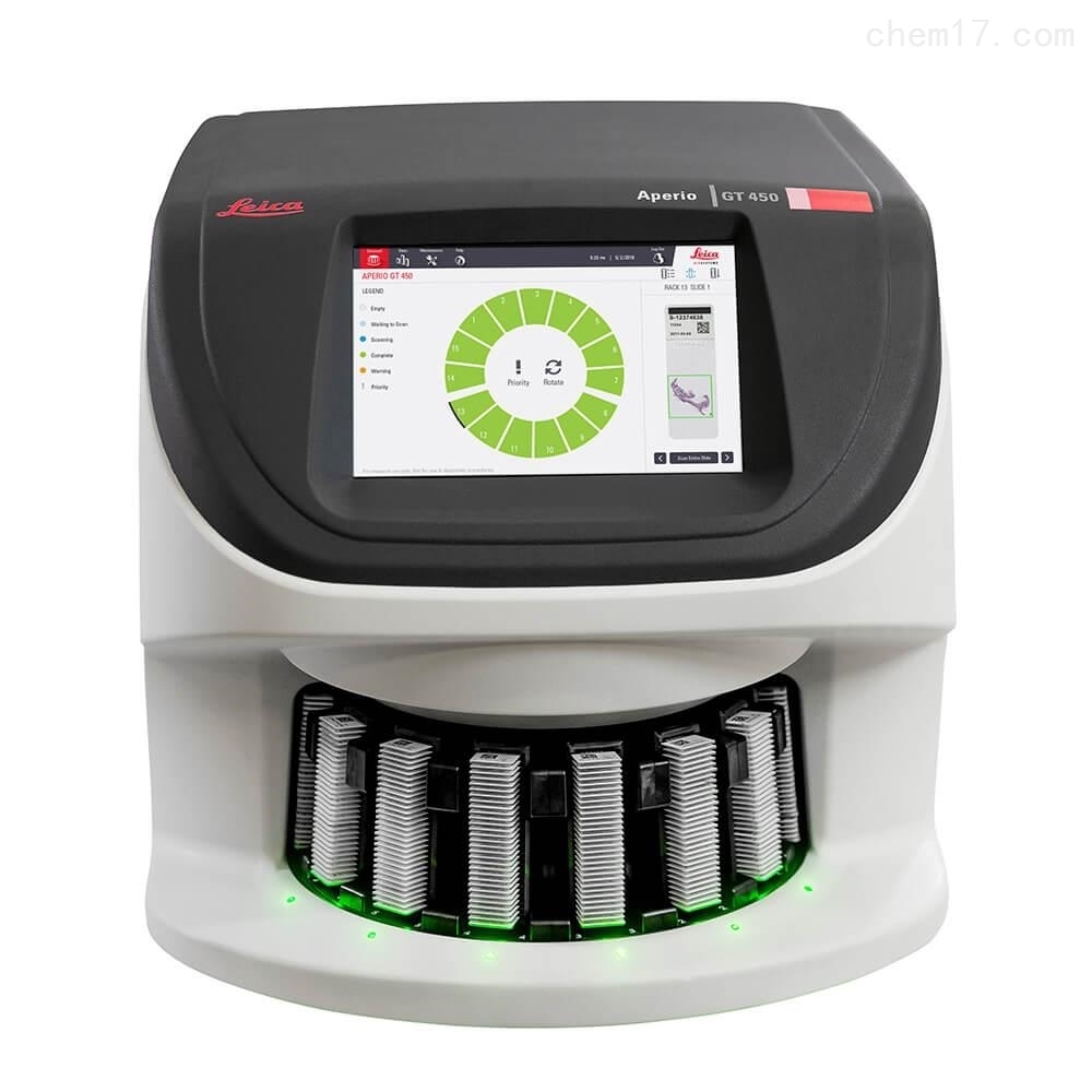
goodslide scannerAdopting high color rendering index (CRI ≥ 95) LED cold light source to avoid color temperature drift of traditional halogen lamps. At the same time, it is equipped with industrial grade CMOS or CCD sensors and undergoes regular white balance calibration. It is recommended to use standard color cards (such as X-Rite ColorChecker) for color benchmarking testing every quarter.
Secondly, software algorithms: color management and ICC configuration files are crucial.
The scanning system should support sRGB or Adobe RGB color spaces and have built-in ICC (International Color Federation) profiles to ensure consistency from capture to display. Some models adopt "multispectral imaging" or "dynamic white balance compensation", which can automatically correct differences in staining batches (such as HE staining being biased towards blue or red).
Thirdly, operation and sample preparation: details determine success or failure.
The glass slide must be clean and free of fingerprints, dust, or sealing bubbles, otherwise it may cause local reflection or color cast;
The sealing agent should be a neutral gum with no spontaneous fluorescence and stable refractive index;
Avoid prolonged exposure to strong light to prevent staining and fading;
Preheat the equipment for 15 minutes before scanning to achieve thermal equilibrium between the light source and sensor.
Fourth, quality control verification: Establish an internal color review mechanism.
Typical HE and IHC stained glass slides can be selected as "gold standard samples" for monthly scanning and comparison to observe whether the cell nucleus is blue purple and the cytoplasm is pink stable. If systematic color deviation is found, the manufacturer should be contacted promptly for optical module calibration.
Only by achieving "what you see is what you get" color reproduction can digital pathology truly replace microscopy as a reliable basis for clinical diagnosis.