-
E-mail
tony.liu@winsafety.com
-
Phone
13925099424
-
Address
Room 301, 3rd Floor, Huangpu Trade Plaza, No. 680 Guangxin Road, Huangpu District, Guangzhou City, Guangdong Province
Guangzhou Mingle Instrument Co., Ltd
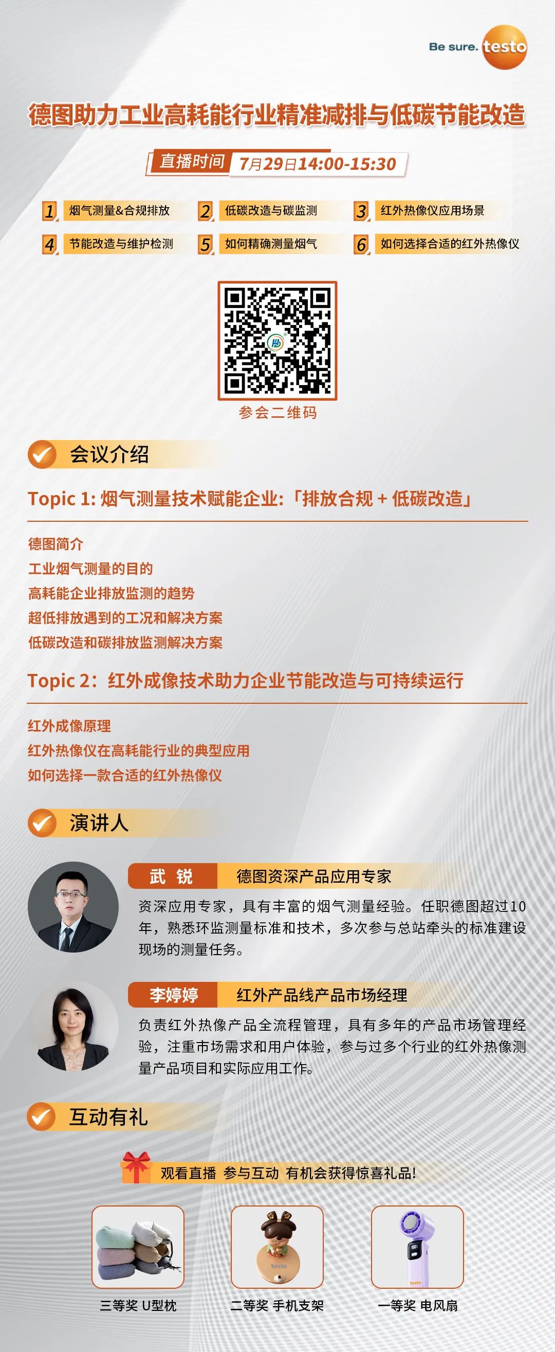
Live Preview | [July 29th] Detu Helps High Energy Consuming Industries with Precise Emission Reduction and Low Carbon Energy Saving Transformation
Date: 2025-07-28Read: 0
Infrared thermography is a passive detection technique that generates temperature field images by detecting the naturally released thermal infrared radiation (wavelength 0.7-1000 μ m) on the surface of living organisms The principle is based on the Stefan Boltzmann law, which states that all objects above absolute zero will continue to emit temperature dependent infrared radiation In the medical field, through the analysis of body surface temperature differences, 0.025 ℃ minor abnormalities can be found early, and the accuracy of breast cancer screening is 81%, and the accuracy of cardiovascular and cerebrovascular disease early warning is over 85% Technically, an uncooled focal plane sensor array is used to convert the radiation signal into an electrical signal, ultimately generating a pseudo color heatmap.
All objects with temperatures above -273 ℃ will emit infrared radiation through molecular thermal motion, and its intensity is proportional to the fourth power of temperature The infrared thermal imaging system consists of three parts: optical lens, detector array, and signal processor:
- Optical lens focuses on the 8-14 μ m wavelength infrared emitted by an object
- The detector array (such as 32 × 24 pixel MLX90640) converts radiation into electrical signals
- The processor uses Planck's radiation law to calculate temperature values and generate color temperature encoded images
In these applications, using thermal imaging technology to make the temperature clearly visible makes your work easier:
In building thermal imagingIn China, the Detu thermal imager can help you detect thermal bridges and structural defects.
In heating engineeringYou can use thermal imaging technology to check the functionality of floor heating systems or non-destructive detection of leaks.
During the maintenance processInfrared thermal imagers can detect wear and tear before the system malfunctions.