-
E-mail
info@excipolar.com
-
Phone
15395560171
-
Address
Building 5, Chuangyan Port, Changzhou Science and Education City, No. 18-50 Changwu Middle Road, Wujin District, Changzhou City, Jiangsu Province, China
Changzhou Puguang Huilian Technology Co., Ltd
Application of near-infrared fluorescence imaging microscope
Date: 2025-11-24Read: 5
Near infrared fluorescence confocal microscopy imaging system
In the fields of biomedical, materials science, optoelectronic devices, and biomedical engineering, target samples (such as deep tissues of living small animals,...)NIR-IIquantum dot/Perovskite micro nano structureCells labeled with biological targeted fluorescent probes often exhibit dynamic evolution and multiple structuresHierarchical andSensitive to complex environmental features, its core performance (such as tissue imaging depth, material luminescence uniformity, probe targeting efficiency) is related to spatial distribution, temporal dynamics, and light-Material interactions are tightly bound together. Traditional single imaging technology can only obtain information from a single dimension, making it difficult to break through Shallow imaging limitations and High background interference The dual bottleneck cannot be fully resolvedNIR-IIThere is a demand for deep and high-resolution imaging in certain frequency bands. The near-infrared two zone fluorescence confocal microscopy imaging system integratesNIR-IIThe advantages of wavelength band, confocal spatial resolution, and in-situ dynamic monitoring function have enabled comprehensive imaging analysis of samples from submicron microstructures to deep tissue layers of several millimeters, which has become a breakthroughNIR-IIThe core technology of bottleneck research in related complex systems.
Any nonNIR-IIThe imaging technology of wavelength bands or non confocal modes both have inherent limitations in deep imaging capability and spatial resolution, which can only reflect partial properties of the sample and cannot fully capture itNIR-IIThe core research needs of the field. For example, although visible light confocal imaging has sub micron spatial resolution, due to the strong scattering and absorption of visible light by biological tissues, the penetration depth is only a few hundred microns, making it difficult to cover deep tissues such as mouse brain and liver;NIR-IFluorescence imaging(700-900 nm)Although the penetration depth has slightly increased, it is still limited byOrganize pan frequency absorptionThe background noise is significant and cannot clearly distinguish the tumor boundary from normal tissue; Ordinary wide fieldNIR-IIAlthough imaging can be utilized Biological Optical Window(1000-1700 nm)Realize millimeter penetration, but lack confocal spatial tomography capability, unable to locate submicron structures in deep tissues (such as capillary branches); Although transient fluorescence imaging can capture luminescence dynamics, it cannot be correlatedNIR-IISome bands have deep spatial distribution —— This requires the use of a near-infrared two zone fluorescence confocal microscopy imaging system “NIR-IIBand low scattering+High resolution confocal microscopy " The synergistic advantage of Z technology fills the key gap in deep and high-resolution imaging, and maximizes its complementarity with other technologies.(Reference:Schmidt et al.,Nat Rev Methods Primers 4, 23 (2024).)
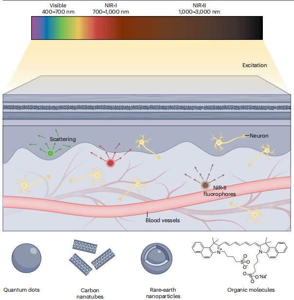
image1. Near infrared two zone fluorescence imaging
In the field of optical information detection, near-infrared two zone fluorescence confocal microscopy imaging systems often integrate in-situ environmental control (temperature, humidity, gas atmosphere, adapted to living biological samples) and multimodal data correlation (synchronous acquisition of fluorescence intensity, spatial tomography images, time-resolved luminescence signals) functions, through high numerical apertureNIR-IISpecialized objective lens focuses excitation light and filters background stray light through pinhole, ensuring a unified space-Synchronized acquisition of three-dimensional fluorescence distribution and dynamic evolution data of samples in depth coordinate system effectively avoids the traditional step-by-step detection Structural misalignment caused by sample displacement and fluorescence quenching caused by environmental changes Wait for the question. Due to the influence of biological tissuesNIR-IIThe scattering and absorption of light are only visible light1/10-1/5And the systemHaving lowPhototoxicity (reduces tissue photodamage), can be analyzed in real-time The Light- NIR-IIprobe-Biological Organization " Cross dimensionalInteraction mechanism —— For example, in live mouse brain imaging, the capillary diameter and blood flow velocity distribution at a depth of several millimeters can be clearly presented; InNIR-IIquantumPoint micro nano structureIn characterization, sub micron localization is possiblelevel areaThe luminous defect. This in-situ fusion strategy not onlyNIR-IIMicro-nano materialsThe study of micro mechanisms such as interface luminescence mechanism and physiological functions of biological tissues (such as tumor angiogenesis) provides direct evidence, and also through Dynamic monitoring+Deep layer chromatography " The combination of technology has laid the core methodological foundation for revealing the cross scale interaction patterns of complex systems, such as the interaction between tumor microenvironment and probes.
The core value of near-infrared two zone fluorescence confocal microscopy imaging system lies not only in “NIR-IIBand advantage+Confocal resolution capability The collaborative application lies more in its generated 'Space'-depth-Time " Deep coupling and intelligent analysis of multidimensional imaging data. This fusion breaks through traditional imaging techniques Shallow Limitations and Resolution-Balance of penetration depth of 'Data Island' Problem, by establishing The sample is emptyInter distribution-Deep organizational response-Dynamic Evolution of Time " The cross scale correlation model has achieved holographic cognition of complex systems —— For example, in tumor diagnosis, the spatial position of the tumor boundary, the blood supply status of deep blood vessels, and the target of the probe can be obtained simultaneouslyTowards integrationEfficiency; InNIR-IIIn material characterization, it is possible to correlate the luminescence intensity of micro regions with the uniformity of crystal structure. Especially in the context of the rapid development of artificial intelligence and deep learning, the high-throughput imaging data generated by this system (such as three-dimensional fluorescence spectra of living tissues, dynamic curves of material micro area luminescence) combined with deep learning algorithms can achieve automatic identification of tumor areas, quantitative analysis of blood flow dynamics parametersNIR-IIIntelligent positioning of material defects will undoubtedly break through the existing limitations Experience driven The imaging and representation paradigm has entered a data-driven stageNIR-IIA new stage of scientific research.
Application case:
1. time-emptyinterval-Multidimensional analysis of deep dynamics and molecular mechanisms of biological tissues using energy
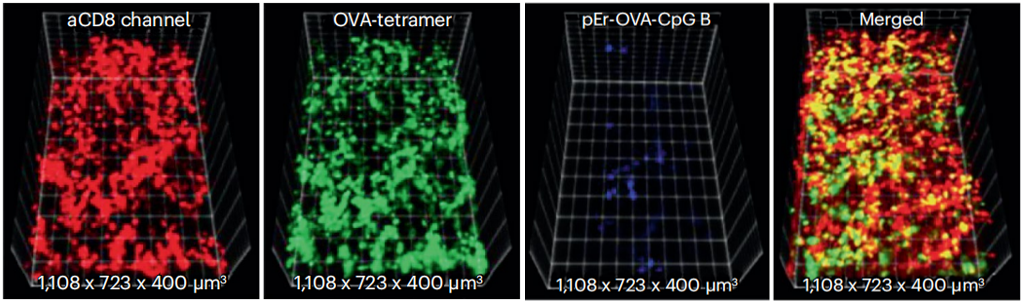
image2. In tumorsCD8+TCell (red), ovalbumin antigen specificityTCells (green) andpernp-ovacase-Bcytosine-phosphoric acid-Guanine(CpG B)Three dimensional nano vaccine (blue)(3D)ReconstructionNIR-IIimage[1]
Furthermore, if an energy axis is introduced on the basis of temporal resolution and spatial imaging(EIf the wavelength of the probe emission is (i.e., the wavelength of the probe emission), then it can be delayed by time(t), spatial location(r)Compared to photon energy(E)Composed of three-dimensional space(t-r-EDraw the emission distribution map of probe signals in phase space, i.eI (t,r,E)Graph. This type of map allows researchers to simultaneously track the temporal evolution of probes (such as blood flow front movement, lymphatic transport time), spatial distribution characteristics (such as tumor targeted aggregation areas, deep vascular branches), and energy adaptation characteristics (such as emission wavelength and...)NIR-IIMatch degree of sub regions), revealing different probes-Selective origin of signal emission in the state of biological interaction. For example, in live biological tissue imaging, different time, space, and energy levelsNIR-IIThe probe signal shows significant differences:FDAapprovedICGBy relying on the tailLaunch extension toNIR-IIa District(1300-1400 nm)Real time localization of sub millimeter in human liver tumor surgeryrank shiftStove (spatial resolution~2 μm)Can track postoperative outcomes in terms of time dimension 24 The dynamic clearance of liver and kidney by probes within hours;NaYbF4:YesRare earth nanoparticles(RENPs)Launch1550 nm NIR-IIbBand signal, still maintained after penetrating the intact skull of miceSNR>10Clearly distinguish the branches of cerebral arteries (red) and veins (blue) in space, match the tissue scattering minimization window in energy, and pass through in time> 20frame/Capture the blood flow fluctuations caused by the cardiac cycle in seconds through dynamic imaging;CH-4T@Protein complex emission1000 nm NIR-IISignal, can be compared with vascular probes in lymphatic imaging(DCNPs,1060 nm)Forming dual color imaging, accurately distinguishing inguinal lymph nodes from surrounding vascular networks in space, and tracking the transport process of probes from intradermal injection to lymph nodes in time(~30Peak in minutes);NIR-IIFluorescence imaging can be used to determineRENPsfor Deep brain tissue imaging priority probe (adapted)NIR-IIbLow scattering window),ICGfor Clinical translation priority probe(FDAApproval attribute), while identifying the attenuation pattern of tissue autofluorescence with increasing wavelength(>1300 nmNegligible) causedSNREnhancement effect.
2. quantum dotprobePreparation andNIR-IIDeep biological imaging applications
Near infrared zone 2(NIR-II,1000-1700 nm)Fluorescence imaging relies onLow organizationThe unique advantages of scattering and low autofluorescence have shown great potential in high-resolution imaging of deep tissues, becoming an important research direction in biomedical imaging. However, the existingNIR-IIFluorescent groups, such as organic dyes, carbon nanotubes, and rare earth nanocrystals, commonly suffer from low quantum yield, poor chemical stability, or insufficient biocompatibility; although PbS Quantum dots(QDs)Has high quantum yield (achievable)60%)But its heavy metals(Pb²⁺)Leakage toxicityS²⁻The optical stability defects caused by easy oxidation severely limit its application in in vivo imaging. In recent years,PbS@CdS core-shell structureQDspass CdS The shell has been preliminarily improvedPb²⁺Leakage issues, and SiO₂andFDAapprovedamphiphilicpolymerPluronic F-127The double-layer coating strategy can further synergistically enhance the chemical stability and biocompatibility of the material——SiO₂The layer provides physical isolation to suppress oxidation and ion leakage,F-127The layer endows with water solubility and anti biological adsorption ability, by constructing PbS@CdS @SiO₂@F-127Nanoparticles(NPs)Expected to break throughNIR-IIfluorescent probe“high brightness-high stability-Low toxicity " The bottleneck that is difficult to balance provides a new solution for imaging deep tissues such as cerebral blood vessels and gastrointestinal tract.
In this study, the target amount was prepared by inverse microemulsion method, surface grafting modification and ultrasonic dispersion processSubpoints, combined with transmission electron microscopy(TEM)Dynamic light scattering(DLS)Steady state/Transient fluorescence spectroscopy, atomic absorption spectroscopy(AAS)And InGaAs Camera imaging system, the system reveals PbS@CdS @SiO₂@ F-127 NPsThe intrinsic optical properties, chemical stability, and biological imaging performance of the material have been found to have high quantum yield(~5.79%Far exceeding most existing onesNIR-IIFluorescent cluster), widthpHScope chemical stability(pH 1-13inside24 hFluorescence changes<30%)Low in vitro and in vivo toxicity(UMUC3cell 10 μg/mLSurvival rate under concentration> 90%), as well as ultra fast in vivo clearance (after gastrointestinal gavage)4.5 hAdvantages such as excretion with feces; These performance improvements are attributed to SiO₂-F-127The synergistic effect of double-layer structure——SiO₂The shell physically isolates oxygen and water to suppressQDsOxidation,F-127ofPEGSegments reduce protein adsorption and enhance water solubility, forming a dense coating between the twoEffective layerblockPb²⁺/Cd²⁺Leakage, at the same timentypeQDsThe interface interaction with the coating layer optimizes the fluorescence emission characteristics.(Reference:Zebibulaet al.,Adv.Funct. Mater. , 32: 2200386 (2022))
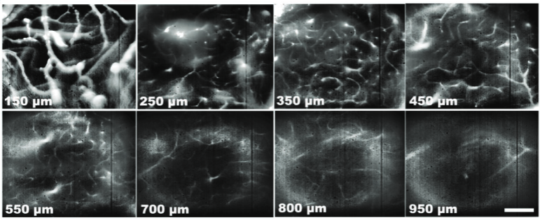
image3. The same mouse cerebral blood vessels captured at different depthsNIR-IIFluorescence image
The experimental results effectively confirmed the monolayer CdS Shell layer PbS The protective effect of nuclear (reducing)Pb²⁺Leakage rate) and SiO₂-F-127The improvement effect of double-layer coating on chemical stability highlights PbS@CdS @SiO₂@ F-127 NPsNIR-IIImaging performance: Its emission peak is located at1155 nm(Adaptation)NIR-IIa Implementation of low scattering window in mouse cerebral vascular imaging 950 μm Clear visualization of deep vascular networks,3DReconstructed images can distinguish the capillaries in the pia mater; In gastrointestinal imaging 80 μg/kgLow dose gavage is sufficient to achieve3-5 mmDeep dynamic tracking of digestive organs(10 minOutline of the stomachThe4.5 hReaching the large intestine and being excreted with feces),7 dThere is no residue in the posterior body. Meanwhile, these results also directly guide the experimental parameter selection of the probe: the excitation wavelength is determined as785 nm(Match) PbS@CdSQDsHigh excitation efficiency), locked imaging detection window1100-1700 nm(Filter out stray light from excitation light); Among them, differentpHThe fluorescence intensity change spectrum under certain conditions provides direct optical evidence for the chemical stability of the double-layer coating, while the dynamic evolution of fluorescence signals in in vivo imaging (such as cerebral vascular blood flow fluctuations and gastrointestinal probe migration trajectories) further verifies the reliability of this quantum dot in deep tissue dynamic monitoring.
3. Biocompatible water-soluble shortwave infrared(SWIR)Launching flowerseliteSynthesis of fluorescence like probes and multi-dimensional breast cancer in vivoDegree moleculeImaging applications
Shortwave infrared(SWIR,900-1400 nm)Imaging relies onLow organizationThe unique advantages of scattering and low autofluorescence show great application potential in the field of biomolecular visualization of deep tissue, especially providing a new direction for accurate imaging of solid tumors such as breast cancer. However, currently available in clinical and scientific researchSWIRFluorescent probes have significant limitations:FDAApproved near-infrared(NIR)Probe indoleeliteGreen(ICG)Although it can be extended toSWIRBand emission, but the excitation wavelength is limited to800 nmThe following only supports monochrome imaging and cannot meet the requirements of multi-target synchronous monitoring; otherSWIRFluorescent groups (such as carbon nanotubes and quantum dots) face problems of poor biocompatibility, insufficient water solubility, or complex synthesis. In recent years, based onICGof π-Conjugated extended flowereliteDyes have attracted attention due to their strong structural tunability and high biological safetyICG-C9andICG-C11By increasing moreJiachuan ChainNumber of double bonds (respectively)ICGmany 1 a、2 aDouble bond) to achieve red shift of emission wavelength toSWIRCore frequency band, and relying onICGThe biocompatibility of the parent nucleus can be optimized for water solubility through surface modification. by constructingICG/ICG-C9/ICG-C11Conjugates of base probes with antibodies, apoptosis markers, or anticancer drugs can synergisticallySWIRImaging advantages and target recognition ability enable multi-dimensional analysis of breast cancer surface receptors, tumor blood vessels and apoptosis processSWIRThe clinical translation of molecular imaging provides new ideas.(Reference:Mahadeva et al.,ACS Appl. Mater. Interfaces. ,16, 17253−17266(2024).)
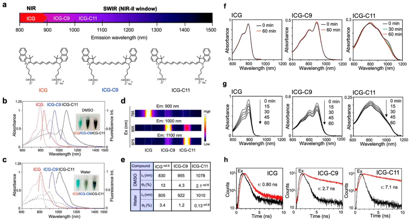
image4. indoleeliteGreen(ICG)TheICG-C9andICG-C11Optical properties ofComprehensive characterization includes absorption spectroscopy, fluorescence spectroscopyFluorescence emission peak and quantum yieldAnd fluorescence emission lifetime
In this study, organic synthesis and steady-state methods were employed/Transient spectral characterization and in vivo imaging technology, the system revealsICG-C9/ICG-C11The intrinsic optical properties and biological imaging performance of the probe have been found to be preciseSWIRBand emission (aqueous phase)1% BSAIn the middle,ICG-C9launch922 nmTheICG-C11launch1010 nm)Excellent biocompatibility(HeLacell 10 MMSurvival rate under concentration> 90%Inside the body 4 Zhou GanshenNormal functional indicators and stable imaging capability(1% BSAMedium laser irradiation60 minphotobleaching rate< 30%)These performance improvements are attributed to π-The wavelength redshift effect caused by conjugate extensionBSAThe composite effect with the probe enhances stability and flowerseliteInherited from the mother nucleusICG生物安全性。
The experimental results effectively confirmedICG-C9/ICG-C11of π-Conjugate extension structure andSWIRCorrelation of emission characteristics (with each increase) 1 aDouble bond, emission wavelength redshift~100 nm), andNHSEsters, MalesacylImines, alkynes, and three types of labeling reagents and antibodies(HerceptinTheErbitux)Theannexin VAnti cancer drugsKadcylaThe efficient coupling ability highlights the multi-color nature of this type of probeSWIRImaging adaptability —— pass785 nm(ICG)The905 nm(ICG-C9)The975 nm(ICG-C11)Differentiated excitation and corresponding band filters(900±25 nm、1000±25 nm、1100±25 nm)Can achieve breast functioncancerHER2/EGFRThree color synchronous imaging of receptors and tumor blood vessels; Meanwhile, this result directly guides the selection of probe combinations for apoptosis imaging(ICG-KadcylaandICG-C9/IC11-annexin V)And wavelength parameters for long-term monitoring(ICG-C9-Kadcylaadopt905 nmStimulate>1000 nmDetection). Among them, steady-state absorption/Fluorescence spectroscopy confirmed the probe-Preservation of optical properties and transient fluorescence after coupling of biomoleculesdecayCurve(ICG-C9lifespan2.7 nsTheICG-C11lifespan7.1 ns)ForSWIRThe time-resolved recognition of signals provides necessary optical evidence, while the signal distribution in live imaging verifies the targeting of probes.