-
E-mail
huogang010@163.com
-
Phone
13911564578
-
Address
Jisheng Villa 19-11, Changping District, Beijing
Beijing Zhongxian Hengye Instrument Co., Ltd
ZEISS LSM 900 MAT Zeiss Material Laser Confocal Microscopy Solution
Date: 2025-09-15Read: 2
The Zeiss LSM 900 MAT confocal laser scanning microscope (CLSM) is a multifunctional imaging system designed specifically for materials research. It can be upgraded with either the upright Axio Imager. Z2m or inverted Axio Observer 7 optical microscope, integrating optical microscopy and confocal imaging capabilities. It can achieve traditional observations such as bright field, dark field, polarization, fluorescence, etc., as well as complete three-dimensional surface morphology analysis and non-destructive measurement through non-contact detection. It is a multidimensional and multi-scale analysis solution for materials science analysis and research. Even with confocal fluorescence and Airyscan, high-resolution fluorescence characterization of biological material samples can be achieved.
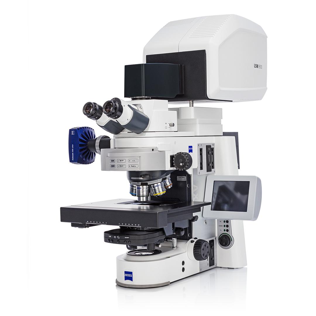
ZEISS LSM900 MAT material laser confocal microscope
1、 Core advantages: technological breakthroughs and system efficiency upgrades
(1) High resolution imaging: from optical principles to hardware guarantees
System based on confocal optical path design: laser controlled by scannerXTheYDirectional scanning of the sample, only information within the focal plane (yellow) can be detected through the pinhole, and information outside the focal plane (red)/The blue dashed line has been filtered; By moving the objective lens away from the sample, a series of optical slices are obtained and stacked, and a height map is generated by combining pixel intensity analysis to achieve three-dimensional imaging.
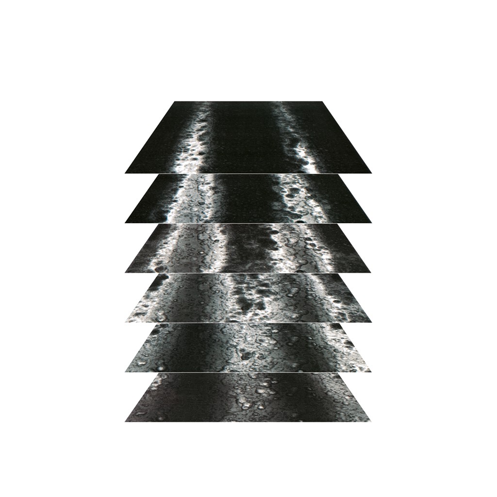
Confocal principle: three-dimensional imaging of the entire sample
In terms of hardware, its core advantage lies inPrecise optical configurationFirstly, it is equipped with a complex chromatic aberration corrected C-Epiplan-APOCHROMAT objective lens, which is optimized for a 405nm laser wavelength, resulting in minimal full field aberration, significantly reduced imaging artifacts and noise - compared to ordinary objectives, it has no edge distortion, a cleaner plane, and more accurate presentation of details; The second is to provide two types of laser configurations to expand the application scope of confocal microscopy: a single channel system (405nm ultraviolet laser, Class 2 laser product) achieves 120nm lateral resolution, and a URGB multi wavelength module (405/488/561/640nm) supports fluorescent dye distribution imaging, suitable for biological material cell observation and other scenarios.
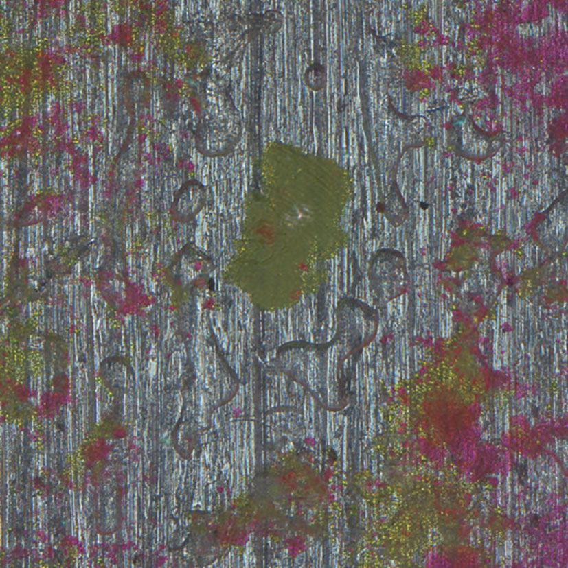
Using different excitation wavelengths to identify color pigments
The final horizontal (XY) resolution reaches 120nm, the minimum height (Z) step is 10nm, and the scanning resolution is up to 6144 × 6144 pixels, which can capture nanoscale details such as metal grain boundaries and polymer interlayer structures.
(2) Flexible imaging: a combination of optical microscope and confocal microscope
The system passes through“Process optimization design”Reduce operational barriers and improve efficiency:
No need to switch devicesComplete wide field positioning and confocal analysis on the same platform, enabling observation of reflected and transmitted light, as well as morphology characterization observation; Using a wide field observation method to locate the sample, facilitating further in-situ analysis using confocal microscopy;
•Save instrument setup timeFor example, the characterization of reflected light from metal metallographic samples can be seamlessly integrated with the observation of transmitted light from rock thin sections;
• Intelligent workflowGuided operation simplifies imaging steps, automated data acquisition supports multi position batch scanning; You can define the region of interest (ROI) in the preview image and only collect the required area to avoid redundant data;
• Full process data controlFrom adjusting imaging parameters to post-processing, it is independently controllable and compatibleZENSeries software—ZEN IntellesisImplement machine learning driven image segmentation and phase discrimination,ZEN ConnectOverlayable management of multimodal experimental images,ZEN Data StorageProvide intelligent data archiving to meet the needs of multi scenario research.
(3) Multi functional extension: covering the full scenario requirements of material research
In addition to basic imaging, the system uses“Module upgrade+Specialized software”Expand application boundaries:
• Quantitative analysis ability: Standard configurationConfoMapSoftware capable of developing roughness(Ra/Rz)Geometric and functional analysis, generating detailed reports containing parameters such as volume, depth, and perimeter; After installing optional modules, 3D Fourier analysis, surface evolution statistics, and grain size analysis can also be performed/Particle counting;
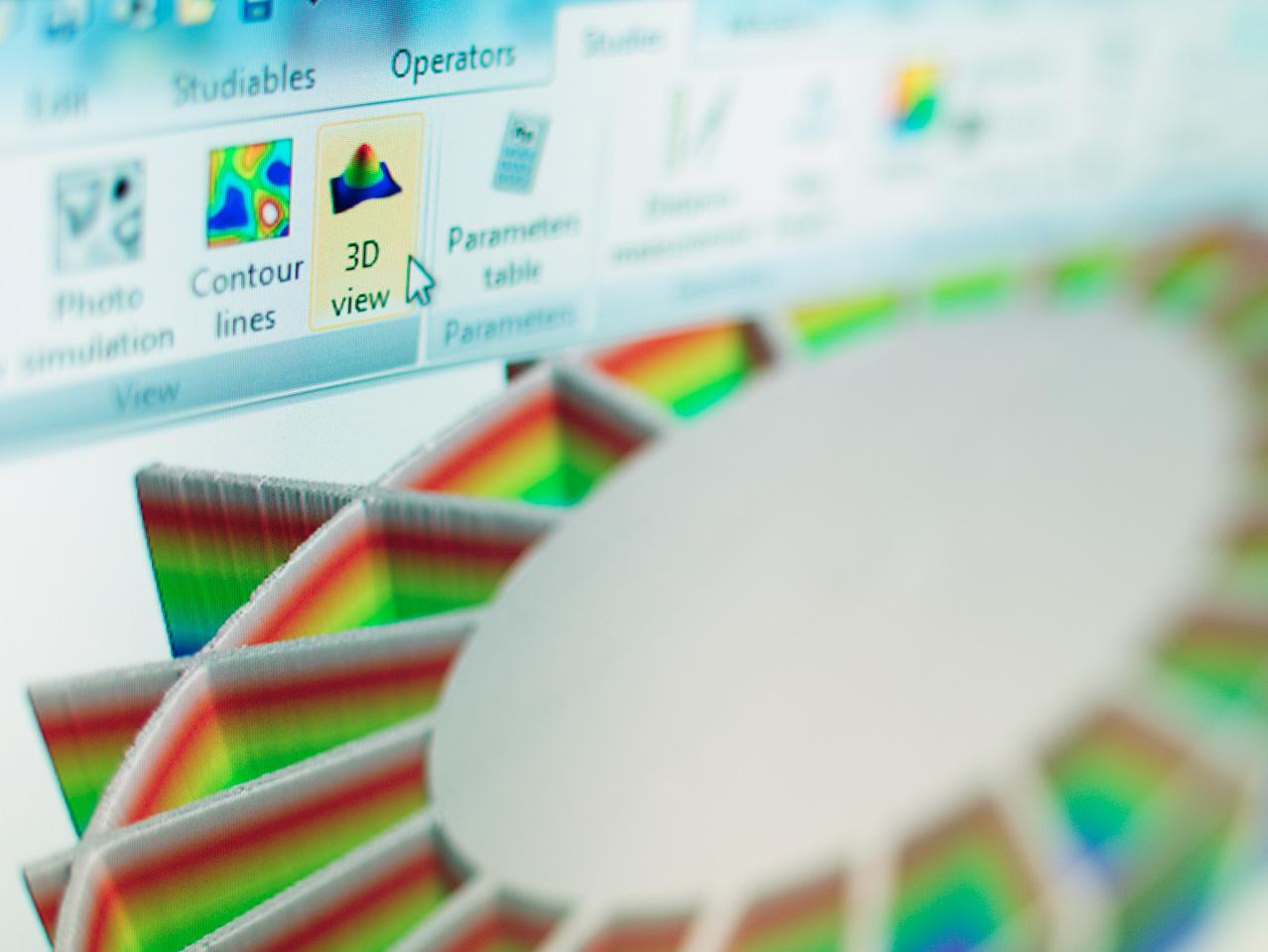
Using ConfoMap for 3D surface inspection
• Non destructive and in-situ testingMeasure the coating without damaging the sample/Thin film thickness, coupled with third-party accessories such as cold and hot tables, stretching tables, etc., can track the dynamic response of materials under environmental changes;
• Optoelectronic combination:Combined with Zeiss electron microscopy, it can achieve multi-scale correlation analysis and integrate microstructure and macroscopic performance data.
2、 Typical applications: from industrial testing to scientific research exploration
(1) Industrial Quality and Failure Analysis
•Analysis of Mobile Phone FailureGenerate texture images, overlay images, and color height maps (image width)1.1mm)Intuitively locate issues such as scratches on the outer shell and deformation of the internal structure;
•Characterization of Metals and AlloysObserve the flatness of the laser polished surface of additive manufacturing alloys and analyze the grain differences between austenite and ferrite near the weld seam of duplex stainless steel (image width)445μm)Assist in optimizing welding processes;
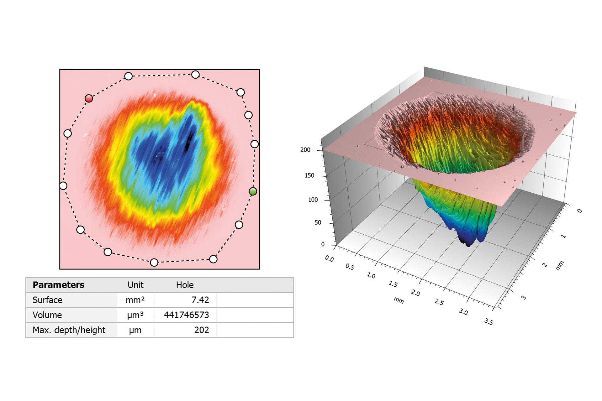
• Wear testMeasure the three-dimensional volume of metal wear holes, derive parameters such as surface area and depth, and quantify the degree of wear.
•Metal testing for material wear and tear.Measurement of the volume of the orifice. A 3D view of a color elevation map. Parameters such as volume, surface, depth, perimeter, and complexity can be exported from the report.
(2) Research on Material Structure and Properties
•Polymers and compositesClear presentation of interlayer interfaces of multi-layer composite polymers, without interlayer signal interference, to assist in multi-layer system design;
• Ceramics and Geological MaterialsCeramic surfaces display micro undulations in color elevation maps; In the study of sandstone porosity, after fluorescence staining4×4Splicing imaging (non-contact measurement), three-dimensional characterization of pore distribution;
•Functional material testingGenerate color 3D views of diffractive optical elements (security feature components) in the document to accurately identify microstructure and anti-counterfeiting features.
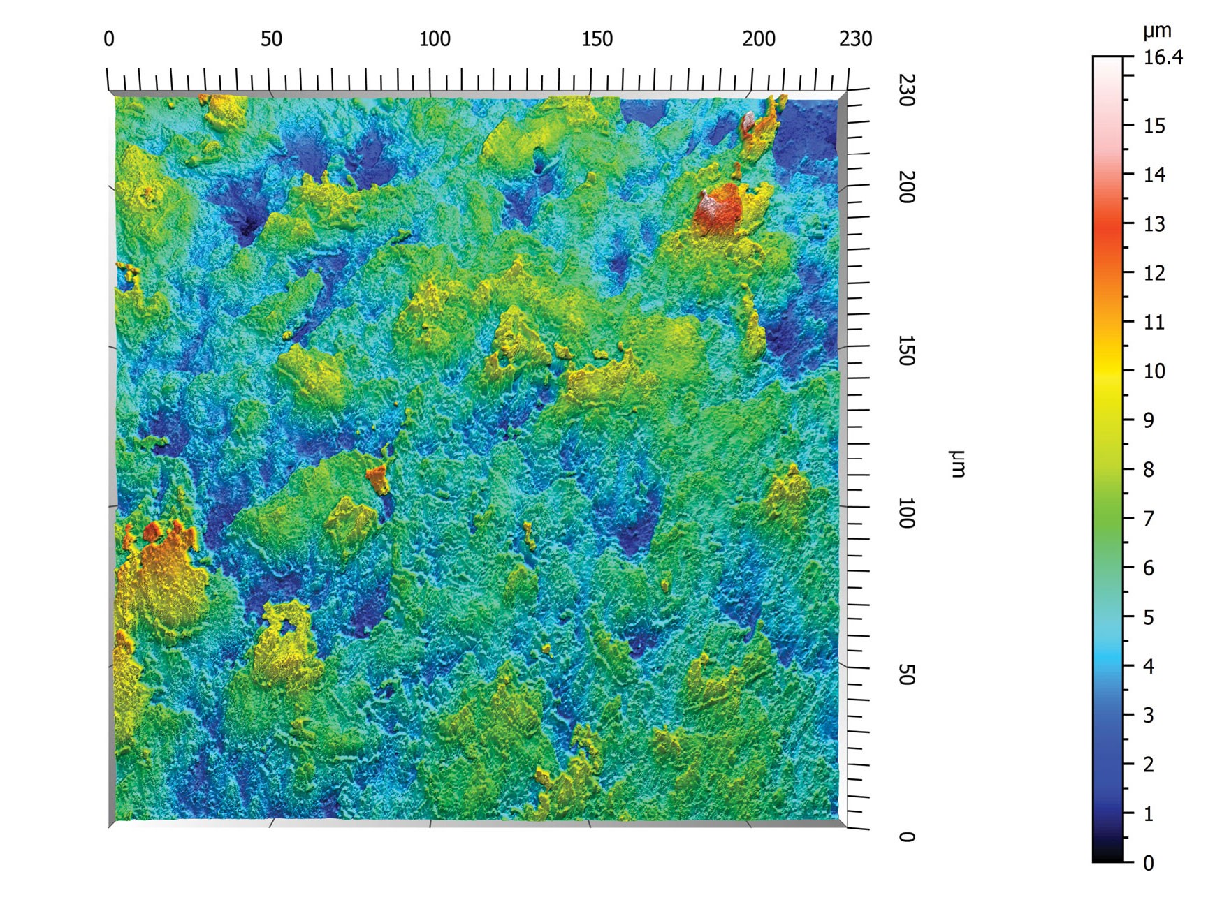
Ceramic surface
(3) Research and development of new energy materials
In the research of perovskite solar cells, the team utilizedLSM 900 MATThe high-sensitivity fluorescence imaging function enables real-time monitoring of the growth process of perovskite thin films. By using multi-color fluorescence labeling technology, the aggregation of iodine vacancies and migration of organic cations at grain boundaries were observed simultaneouslyAIThe auxiliary analysis established a quantitative relationship between defect density and photoelectric conversion efficiency. Improved efficiency of batteries prepared after optimizing the process8%The system's fast spectral imaging capability can 10 Complete the defect distribution of the entire battery within minutesMapping.
(4) Industrial Quality Control
A certain automobile manufacturer has introducedLSM 900 MATConduct quality inspection of aluminum alloy laser welding joints. Combining the reflection light imaging mode of the systemAIDefect recognition algorithm successfully detects microcracks and porosity defects that are difficult to detect by traditional methods, with a minimum recognizable size of1μm. Through3DReconstruction technology accurately measures the volume, distribution density, and spatial orientation of defects, providing quantitative basis for optimizing welding process parameters and improving the welding qualification rate after application15%In the hydrogen energy industry, this system can inspect the surface structure and roughness of electrolytic cell plates, while observing the integrity of the diaphragm fiber structure to ensure the performance of key components.
(5) Geological material analysis
LSM 900 MATThe reflected light scanning mode can reconstruct the three-dimensional structure of rocks, locate the position and depth of pores, and combine fluorescence labeling to locate the remaining oil position, helping to evaluate the difficulty of mining and develop reasonable plans. Its sufficiently large sample storage space supports the morphology and size analysis of various geological samples, and the multi-channel acquisition function of light and heavy components provides key microscopic data for energy exploration.
3、 Future outlook: Empowering innovation in multiple fields
With the development of materials science towards multifunctionality and composites,LSM 900 MATContinuously expanding application boundaries through continuous software upgrades and module extensions. In the field of sustainable materials research and development, its mild imaging conditions make it possible to observe the in-situ degradation process of biobased composite materials; Provide micro characterization support for quality control of key components in hydrogen electrolysis cells in the field of new energy. From laboratory basic research to production line quality control,The performance and flexibility of LSM 900 MAT have become characterization tools in the field of materials science. It not only helps researchers uncover the microscopic mysteries of materials, but also accelerates the transformation of scientific research results into industrial applications through standardized analysis processes and quantitative data. With the support of Zeiss optics technology, LSM 900 MATWill continue to leadThe development of material microscopic characterization technology provides a continuous driving force for innovation in new materials and processes.
References
[1] Zeiss Corporation ZEISS LSM 900 Material Research and Application Guidelines
[2]Zeiss Corporation ZEISS LSM 900: Airyscan 2 super-resolution imaging technology [R]. Carl Zeiss Microscopy GmbH, 2024
[3] Application of confocal microscopy technology in the study of perovskite solar cells [EB/OL]. Materials Science Network, 2025
[4] Zeiss Industrial Microscope Solution. Metal processing, 2025