-
E-mail
selina@cy-tech.com.cn
-
Phone
13426082940
-
Address
1-2F, Building C, Xisanqi Cultural and Technological Park, No. 27 Xixiaokou Road, Qinghe Xiaoying, Haidian District, Beijing
Beijing Saitaike Biotechnology Co., Ltd
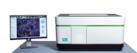
The use of PE high connotation imaging analysis system has significant implications in various aspects
Date: 2025-10-20Read: 0
The use of PE high connotation imaging analysis system has significant implications in various aspects, specifically reflected in the following key areas:
1. Improvement of scientific research efficiency and accuracy
Automated operation optimization process: The system adopts automated microscope technology, which can automatically adjust the focal length, aperture, and light source intensity to achieve fast and accurate sample scanning. This highly automated design reduces the need for manual intervention, not only improving the reproducibility of experiments, but also significantly reducing operation time, enabling researchers to more efficiently analyze large amounts of samples.
Multi parameter quantitative analysis enhances data depth: Through multi-channel fluorescence detection function, the system can simultaneously label and detect multiple cellular components or molecular markers. Combined with advanced image processing software, precise measurement and quantitative analysis of cell morphology, quantity, fluorescence intensity, etc. can be carried out, providing a more comprehensive data dimension than traditional methods, thereby deepening the understanding of biological processes.
Intelligent algorithms ensure the reliability of results: The built-in image analysis algorithm can automatically recognize complex cell features, reducing subjective errors caused by human judgment. Especially in situations of long-term operation or environmental changes, the automatically calibrated optical path design effectively reduces the impact of optical distortion, ensuring the stability and consistency of image quality.
2. PE high connotation imaging analysis system accelerates drug development and innovation breakthroughs
High throughput screening drives new drug discovery: In the early stages of drug development, the system supports large-scale cell level toxicity testing, proliferation inhibition experiments, and migration ability evaluation of candidate compounds. This high-throughput characteristic allows researchers to quickly eliminate invalid molecules, focus on potential lead compounds, and significantly shorten the research and development cycle.
Dynamic process visualization analysis mechanism: With the help of high-resolution imaging technology, researchers can observe real-time changes in intracellular signaling pathways, protein localization and transfer, and dynamic responses of subcellular structures under drug action. These intuitive data provide strong evidence for revealing the mechanism of drug action, which helps optimize compound structures and administration regimens.
Biomarker mining helps precision medicine: By systematically comparing cell phenotypes under different treatment conditions, the system can help identify specific biomarkers related to therapeutic efficacy. These findings not only enrich the database of disease diagnostic indicators, but also provide scientific basis for the design of personalized treatment plans.
3. Cognitive innovation in basic life sciences
Upgraded exploration tools for the microcosm: from cell cycle regulation to differentiation fate determination, and then to the initiation of apoptosis programs, the system is able to capture subtle differences in these basic life activities. For example, through single-cell detection capabilities, scientists are able to decipher rare subpopulation features in heterogeneous populations, breaking through the limitations of previous population average data.
Hypothesis for disease model construction verification: It has become possible to establish an in vitro disease model using primary cells derived from patients. Researchers can simulate the cellular interaction network in pathological environments, verify theoretical hypotheses about the pathogenesis, and lay the foundation for subsequent clinical translational research.
Interdisciplinary cross fusion platform: The massive standardized dataset generated by this system promotes the application of computational biology, enabling machine learning algorithms to extract hidden pattern patterns from it. This data-driven research paradigm is changing traditional research models and giving rise to new scientific discoveries.
4. Standardization and repeatability guarantee of PE high connotation imaging analysis system experiment
Strict control of experimental conditions: The built-in environmental control system of the system maintains constant parameters such as temperature and humidity, ensuring that each experiment is conducted under the same conditions. The standardized operation process reduces inter batch variability, improves the comparability and credibility of results.
Complete traceability of data management system: The supporting software platform realizes the full process digital management from image acquisition to data analysis. The processing history and parameter settings of each sample are recorded in detail, facilitating subsequent audits and reproducing experiments, in line with the transparency requirements of modern scientific research.
Collaboration and sharing promote knowledge dissemination: Open data formats and remote access support enable easy sharing of research results between different laboratories. This interconnectivity has accelerated academic exchanges within the field and promoted the progress of collective wisdom.
Last Article: Key technical methods of single-cell immune analyzer
Next Article: The flexibility and multifunctionality of 3D perfusion culture