-
E-mail
info@tvscope.cn
-
Phone
15261811715
-
Address
Super dimensional scenery
Beijing Chaoweijing Biotechnology Co., Ltd
Neuroscience 'Color Vision': Peking University team successfully develops multi-color miniaturized two-photon microscope
Date: 2025-08-25Read: 4
Recently, the team led by Cheng Heping and Wang Aimin from Peking University, together with the team led by Wu Runlong from Beijing University of Information Science and Technology, successfully developed a multi-color miniaturized two-photon microscope (FHIRM-TPM 3.0), which enables multi-color, deep brain, and cross scale neuroimaging in freely moving mice, providing a new tool for decoding complex brain functional mechanisms. The related research paper "A Versatile Miniature Two Photon Microscope Enabling Multi Color Deep Brain Imaging" was published online in Nature Methods and was recommended by the Research Briefing during the same period.
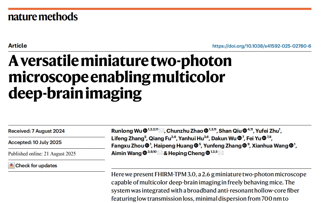
Screenshot of the online publication of the paper
Ten years of sharpening the sword, creating four generations of 'brain science weapons'
The brain is the most complex and intricate "microcosm" in nature, consisting of billions of neurons and trillions of synapses working together. How to accurately capture the dynamic changes in neuronal and synaptic activity in freely moving living organisms is a core challenge in neuroscience research.
In 2014, Academician Cheng Heping led the national major instrument and equipment project "Ultra high spatiotemporal resolution miniaturized two-photon in vivo microscopy imaging system", which took more than ten years to complete four technological iterations:
01 Nature Methods, two thousand and seventeen
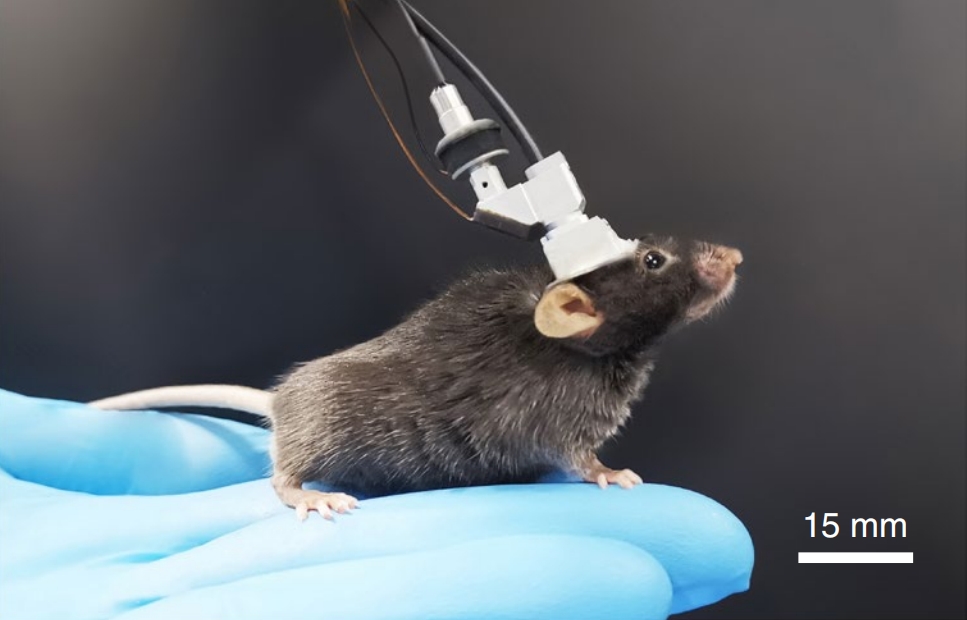
In 2017, the first generation 2.2g miniaturized two-photon microscope was developed, which achieved clear and stable functional imaging of freely moving mouse synapses for the first time.


02 Nature Methods, two thousand and twenty-one
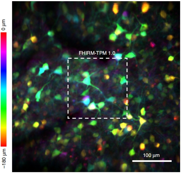
The second generation system in 2021 has expanded the field of view by 7.8 times and has three-dimensional imaging capabilities.
03 Nature Methods, two thousand and twenty-three
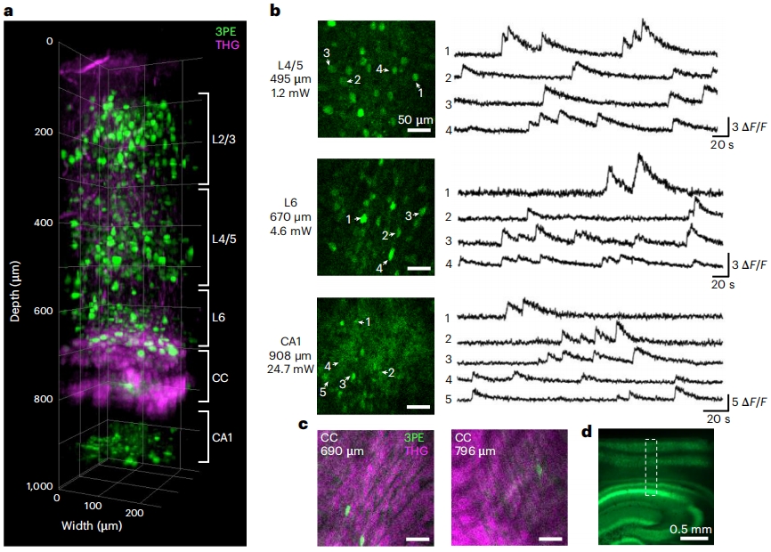
The third generation system in 2023 adopts the principle of three photon fluorescence imaging to achieve non-invasive observation of the deep hippocampus region.
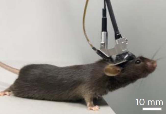
04 Nature Methods, two thousand and twenty-five
The new generation of miniaturized two-photon microscopes has made significant progress in multi-color excitation, deep brain observation, and cross scale imaging.

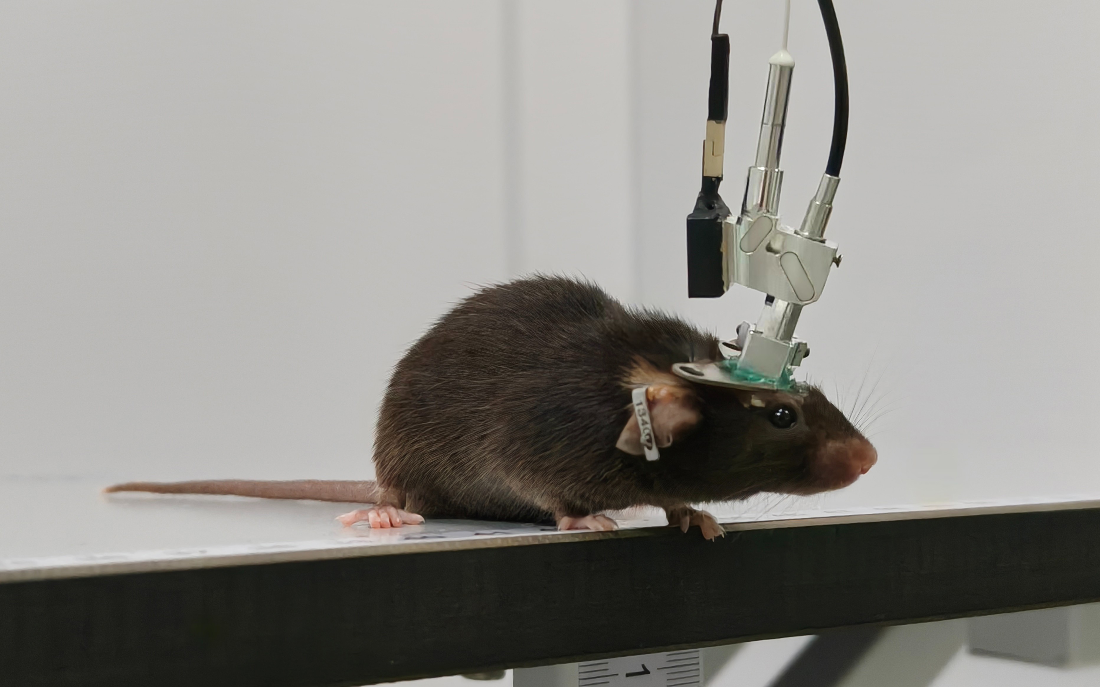
Three color live streaming of brain activity
Previously, hollow photonic crystal fibers only supported laser transmission at a single wavelength, limiting their ability for multi-color imaging. The team has developed 700-1060 nanometer ultra wideband anti resonant hollow fiber, which enables multi wavelength femtosecond pulse laser transmission with low loss, low dispersion, and low bending loss. In the study of Alzheimer's disease model mice, three color synchronous imaging of neuronal calcium signals, mitochondrial (organelle) calcium signals, and brain amyloid plaque deposition was obtained, and mitochondrial calcium dynamics abnormalities in the early stages of the disease were discovered.
Video 1. Three dimensional reconstruction images of neuronal calcium signals (red), mitochondrial calcium signals (green), and amyloid plaques (blue) in Alzheimer's disease model mice
Deep brain imaging: Perspective of 850 micron cortex
By precision optical design for color and spherical aberration correction of the entire system, and optimizing the optical system design, the new generation system has obtained neuronal calcium signals and structural imaging at a depth of over 850 microns in the mouse cerebral cortex, which is more than three times deeper than previous miniaturized two-photon microscopy imaging.
Video 2. Thy1 YFPH transgenic mouse cerebral cortex 854 micron depth 3D imaging reconstruction
Vision switching: achieving cross scale imaging
The team has designed three rapidly switchable autofocus objectives with high resolution, high performance, and large field of view, achieving seamless conversion between large field of view observation and high-resolution fine imaging, and realizing imaging across 10 times the scale from hundreds of micrometers to millimeters.
Video 3. (Left) FHIRM-TPM 3.0 high-resolution probe achieves dual color imaging reconstruction of spatial distribution of neurons in the motor cortex (red) and mitochondria within neurons (green); (Right) FHIRM-TPM 3.0 large field probe achieves imaging of 450 neurons in the prefrontal cortex of freely moving mice
research team
The co first authors of this paper are Professor Wu Runlong from Beijing University of Information Science and Technology (then a specially appointed associate researcher at the School of Future Technology, Peking University), Assistant Researcher Zhao Chunzhu from the School of Future Technology, Peking University, and PhD student Qiu Shan; The co corresponding authors are Cheng Heping and Wang Aimin from Peking University, and Wu Runlong from Beijing University of Information Science and Technology. The project has received funding support from the Major Project of Science and Technology Innovation 2030- "Neuroscience and Brain like Research", the National Natural Science Foundation of China, the National Major Scientific Research Instrument Development Special Project, and the Key R&D Program of the Ministry of Science and Technology.
[Reference]
Wu, R., Zhao, C., Qiu, S.et al. A versatile miniature two-photon microscope enabling multicolor deep-brain imaging.Nat Methods (2025).https://doi.org/10.1038/s41592-025-02780-6