-
E-mail
sales@always.ltd
-
Phone
15300515116
-
Address
Building 3, No. 800 Jiuxin Road, Songjiang District, Shanghai
Aoying Detection Technology (Shanghai) Co., Ltd
Customer Literature | Research on Micro nano Hemostatic Materials Based on Beetle Pulse Jet Biomimetic Structure
Date: 2025-09-24Read: 0
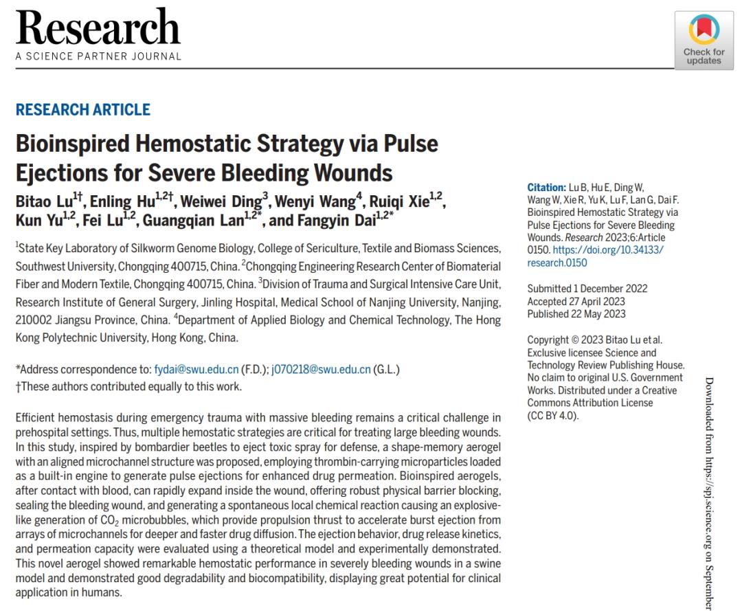
Southwest University research team in internationally renowned journal《Research》An important research achievement has been published. Inspired by the pulse jet mechanism of the Bombardier beetle, this research designed a shape memory aerogel with directional microchannels. Through the micro focus CT imaging system and multi-scale experiments, its hemostatic performance and mechanism were systematically studied.The first author of this study is Professor Lu Bitao.
Research Abstract
Inspired by the defense mechanism of Bombardier beetle ejection, a pulse jet bionic gel was designed to stop serious bleeding wounds. This structure constructs a silk fibroin nanofiber/chitosan aerogel with parallel microchannels by directional freeze casting. It loads CaCO ∨ and tranexamic acid (TXA-NH ∨) as "built-in engines", and uses blood triggered CO ₂ microbubble pulse jet to enhance drug penetration. Combined with shape memory characteristics and physical barrier function, the aerogel rapidly expands and seals the wound after contacting with blood, and accelerates the diffusion of hemostatic agent through gas power. with the help ofMicro focus CT (AX-2000CT)The 3D non-destructive tomography of the aerogel clearly showed the parallel array structure (15-35 μ m wide) and the overall honeycomb porous morphology (52% porosity) of the bionic microchannel, providing key structural data support for the effectiveness of the microchannel structure design and subsequent performance analysis. Through theoretical models, high-speed cameras, and animal experiments, it has been verified that under the optimal parameters, the drug release rate exceeds 50% within 60 seconds, and the hemostasis time in the pig femoral artery hemorrhage model is only 3.5 minutes. The research provides new ideas for hemostasis of complex deep wounds and demonstrates the potential application of biomimetic structures in the field of emergency medicine.
Performance testing methods
In vitro characterizationScanning electron microscopy (SEM) observation of microchannel structure,Micro focus CT (AX-2000CT)Analyze porosity and 3D structure, and evaluate hydrophilicity using a contact angle measuring instrument.
Materials and Research Methods
Analysis of Microchannel Structure
CT imaging shows that the aerogel has a honeycomb porous structure (porosity 52%), the width of parallel microchannels is 15-35 μ m, and the inner wall fibroin nanofibers form an interwoven network, which significantly improves the liquid absorption rate (1.5 × 10 ⁻ ³ m/s). The loaded CaCO ∝/TXA-NH ∝⁺ is evenly distributed in the microchannels, with a gelatin cap coverage rate of>80%, ensuring efficient gas accumulation.

(C) Longitudinal section of GCSF-3/CT (pink pseudocolor represents calcium carbonate (CaCO))₃)The blue pseudocolor represents ammonium tranexamic acid (TXA-NH)₃⁺))And corresponding element distribution images. (D) Top and bottom views of GCSF-3/CT. (E) Micro CT images of GCSF-3/CT.

(B) Display images of the rapid jet phenomenon triggered by water in GCSF-3/CT at high frame rates.
research findings
Adequate hemostasis of severe bleeding wounds is very challenging. The bionic gel prepared in this study can rapidly expand and compress the wound after contacting with water/blood, and accelerate the spread of payload through microbubble power. The drug release rate exceeds 50% within 60 seconds, which shows better hemostasis effect than existing materials in four bleeding models (including pig femoral artery massive hemorrhage), and has good biocompatibility.Micro focus CT (AX-2000CT)It played a key role in the research: it accurately characterized the microchannel structure, porosity and 3D morphology of aerogel through nondestructive 3D tomography, providing direct evidence for verifying the effectiveness of bionic design, analyzing the relationship between structure and hemostatic performance, and providing quantitative basis for material optimization.
original literature
[1]LuB,HuE,DingW,etal. BioinspiredHemostaticStrategyviaPulseEjectionsforSevereBleedingWounds[J]. Research,2023,6:Article0150.DOI:10.34133/research.0150
Paper application equipment

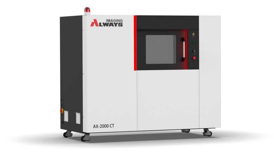
The MicroCT equipment used in this study is a high-precision micro nano CT imaging system developed and produced by Aoying——AX-2000CTThe system is specially designed for biomedical and material science research, and has high precision imaging, fast scanning efficiency, economic cost and diversified application scenarios. It can conduct nondestructive 3D tomography for various samples such as gas mimicking gel, nanofiber scaffold, biological composite materials, small metal components, geological cores, etc., providing key non-destructive digital imaging technology support for bionic structure analysis and performance optimization.
Last Article: The detailed operation process of the micro nano CT machine is as follows
Next Article: How is the micro focus CT machine operated?